
- Title
-
In vivo screening and discovery of novel candidate thalidomide analogs in the zebrafish embryo and chicken embryo model systems
- Authors
- Beedie, S.L., Rore, H.M., Barnett, S., Chau, C.H., Luo, W., Greig, N.H., Figg, W.D., Vargesson, N.
- Source
- Full text @ Oncotarget
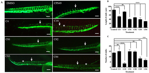
The effect of novel thalidomide analogs on vessel length. fli1:EGFP zebrafish were incubated with vehicle or compounds for 24 hours, at which point vessel outgrowth and number were measured. A. Images of zebrafish exposed to the vehicle or test compounds. Zebrafish with the vehicle (0.1% DMSO) show normal patterning of the ISVs, zebrafish exposed to CPS49 show reduction in vessel outgrowth (white arrow), loss of vascular connectivity and a decrease in the number of forming blood vessels. Treatment with C4 (1.5 µg/mL), C18 (50 µg/mL), C81 (50 µg/mL), C82 (5 µg/mL), C83 (20 µg/mL) and C84 (50 µg/mL) show reduction in vessel out growth (white arrow) and in some cases loss of vessel (white asterisks). B. Stalk length & C. stalk number are reduced. Compounds are shown at the lowest concentration producing an effect. The data represent the mean + the standard error of the mean. |
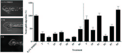
The effect of thalidomide analogs on the inflammatory response to wound healing. A. Zebrafish (72 hpf) embryos transgenic for a fluorescently tagged neutrophil marker Tg(MPO::EGFP)114 were fin clipped to induce an inflammatory response and exposed to vehicle or a test compound. Broken line indicates the tail fin with cut and area of the wound site. B. The percentage of neutrophils within the wound site was quantified and shows the extent of the inflammatory response to injury. The data represent the mean + the standard error of the mean. |

Defects seen in zebrafish and chicken embryos following thalidomide analog treatment A. Embryos with exposure to a vehicle control show normal development of the eye (e), and B. pectoral fins (pf). Example of an anti-angiogenic compound (C4, Figure 1) causing C. microopthalmia (white arrow; seen in list the compounds) and D. malformation in fin development (white arrow). Exposure to an anti-inflammatory compound (C2, Figure 1) resulting in E. normal eye development and F. fin development. G. Eye diameter and H. fin length were quantified and show anti-angiogenic compounds causing reductions in eye diameter (C4, C18, C82, C83, C84) and pectoral fin length (C18, C81, C82). Compounds of interest were screened in chicken embryos at HH stage 17-18 (Day 2.5 in embryonic development). I. Untreated, control images of an embryo in ovo with normal vascular patterns at HH stage 23 (Day 3.5); K. ex ovo; M. eye O. forelimb and Q. hindlimb showing cartilage patterning (at day 9 of development). Typical examples of compound treated embryos: J. anomaly in vasculature (white arrow) and necrosis (black arrow) of the YSM in a chicken embryo following treatment with C23 (Supplementary Table S1). L. embryo exhibiting microopthalmia (asterix), limb reduction (white arrow) and hemorrhaging throughout the body following treatment with C80 and (N) C81 (Figure 1) treated embryo with growth reduction and hemorrhaging throughout head (white arrow). P. C11 treated limb with reduced elements (Asterisks represents a limb reduction defect). R. Missing digits and reduced length of limb cartilage elements (Treated with C11, Supplementary Table S1). Scale bar represents 1000 µm. |