
- Title
-
Conserved and highly expressed tRNA derived fragments in zebrafish
- Authors
- Soares, A.R., Fernandes, N., Reverendo, M., Araújo, H.R., Oliveira, J.L., Moura, G.M., Santos, M.A.
- Source
- Full text @ BMC Mol. Biol.
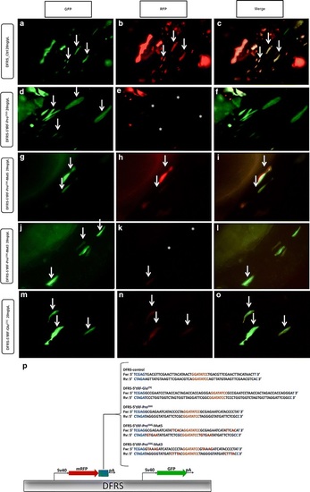
Silencing ability of tRFs using a dual reporter plasmid. Endogenous 5′tRF-ProCGG has silencing ability. Embryos injected with 20 ng/µL of DFRS control plasmid (a, b, c), show GFP and mRFP signal, while the RFP signal is lost after microinjection of 20 ng/µL DFRS-5′tRF-ProCGG plasmid (d, e, f). 5′ portion of 5′tRF-ProCGG is necessary for target silencing, as mutations in the reporter that affect binding at the 5′ end (DFRS-5′tRF-ProCGG_Mut5) result in recovery of RFP fluorescence (g, h, i) when compared to non-mutated reporter (d, e, f). Mutations that affect binding of the 3′ end of the tRF do not affect silencing ability as RFP is repressed (j, k, l), similarly to the non-mutated reporter. There is only a slight decrease in RFP signal after microinjection of 20 ng/µL DFRS-5′tRF-GluCTC (m, n, o) plasmid, indicating that the endogenous 5′tRF-GluCTC does not have trans-silencing ability. Arrows indicate cells containing both GFP-reporter and mRFP-sensor. Asterisks indicate muscle fibers that lost mRFP fluorescence. Orientation of embryos: caudal, left; ventral, up ×20 magnification. p DFRS plasmid scheme. The DFRS plasmid bears two fluorescent proteins, namely GFP (green) and mRFP (red), controlled by SV40 promoters. The mRFP contains a 3′UTR cassette (blue box) complementary to the tRF of interest. The sequences inserted in the DFRS to obtain the different reporters (DFRS-control, DFRS-5′tRF-GluCTC, DFRS-5′tRF-ProCGG, DFRS-5′tRF-ProCGG-Mut5 and DFRS-5′tRF-ProCGG-Mut3) are depicted. Restriction sites are shown in blue, EcoRV cleavage site is shown in orange and the mutated nucleotides are highlighted in red |
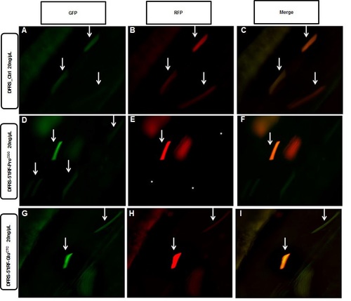
Sensor plasmid at 72 hpf. At 72 hpf GFP and RFP expression is equivalent after the injection of the control reporter (A, B, C). Silencing by endogenous 5′tRF-ProCGG is still present at 72hpf (D, E, F), as RFP expression is repressed. Endogenous 5′tRF-GluCTC does not efficiently silence putative targets even at 72hpf, as no alterations in RFP expression is observed when compared to GFP expression (G, H, I). Arrows indicate cells containing both GFP-reporter and mRFP-sensor. Asterisks indicate muscle fibers that lost mRFP fluorescence. Orientation of embryos: caudal, left; ventral, up. 20× magnification. |