
- Title
-
Mapping of Brain lipid binding protein (Blbp) in the brain of adult zebrafish, co-expression with aromatase B and links with proliferation
- Authors
- Diotel, N., Vaillant, C., Kah, O., Pellegrini, E.
- Source
- Full text @ Gene Expr. Patterns

Blbp distribution in the brain of adult zebrafish. A-L: Blbp (red) immunohistochemistry on paraffin brain sections of adult zebrafish. DAPI counterstaining (blue) allows the visualization of cell nuclei. Blbp staining is detected in radial glial cells along the ventricular layers of the dorsal telencephalic area at the junction with the olfactory bulbs (A), in the ventral and dorsal nuclei of the ventral telencephalon, and in the dorsomedian, the dorsolateral, the dorsal and the posterior zone of the dorsal telencephalic area (B-D). In the RMS region (see asterisks in B), no Blbp staining was detected. Blbp-positive radial glial cells are also observed in the preoptic area and the hypothalamus (D-F). Strong Blbp staining is notably observed in cells surrounding the lateral and posterior recess of the hypothalamus (H). Radial glial cells from the periglomerular gray zone of the optic tectum are also positive for Blbp and extend long cytoplasmic process to the periphery of the brain (G and J, see arrowheads in J), in the periventricular pretectal nucleus (G). Blbp staining around the torus longitudinalis, the torus semicircularis, in the nucleus of the medial longitudinal fascicle (NMLF) and along the vascular lacuna of area postrema (Vas) correspond to Blbp-positive fibers (G and I). Few positive cells are observed in the valvula of the cerebellum and the cerebellum (I and K) and lining the rhombencephalic ventricle (K-L). Arrowheads highlight some long radial cytoplasmic processes crossing the brain parenchyma to reach the periphery of the brain (C, F and J). Arrows show the end of radial glial process at the pial surface (A, D, F and H). M-O: Blbp (red) and HuC/D (green, neuronal marker) double immunohistochemistry reveals that Blbp is not expressed in mature neurons. Arrowheads indicate Blbp positive radial glial cells that do not express the HuC/D neuronal marker. Bar: 16 µm (E, J, M, N and O); 50 µm (C, F, G and L); 100 µm (A, B, D, H, I and K). |
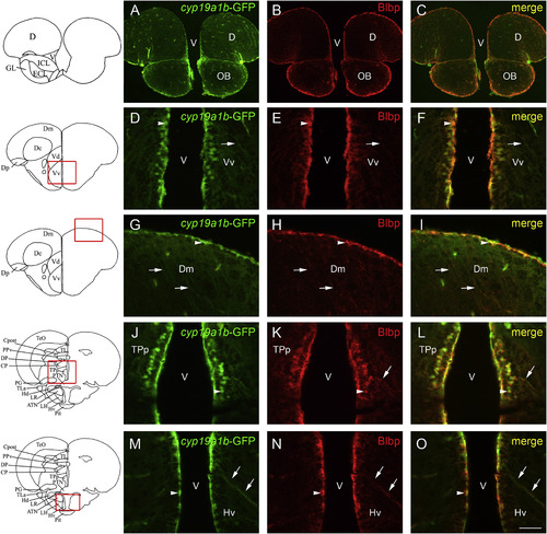
cyp19a1b-GFP and Blbp co-expression in the brain of adult zebrafish. A-O: Blbp (red) immunohistochemistry on cyp19a1b-GFP (green) transgenic zebrafish line shows that most radial glial cells co-express Blbp and GFP. Co-expression is obvious at the junction between the telencephalon and the olfactory bulbs (A-C), in the subpallium (D-F), and in the pallium (G-I). In the anterior part of the periventricular nucleus of the posterior tuberculum, GFP and Blbp are co-expressed (J-L) as well as in the anterior region of the hypothalamus (M-O). Arrows point to radial glial processes co-expressing GFP and Blbp, while arrowheads highlight co-expression in soma. Bar: 25 µm (D-O); 100 µm (A-C). |
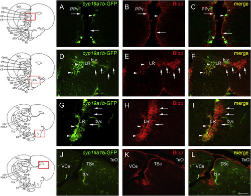
cyp19a1b-GFP and Blbp show differences of expression in more caudal regions of the brain of adult zebrafish. A-L: Blbp (red) immunohistochemistry on cyp19a1b-GFP (green) transgenic zebrafish line show discrepancies in expression of AroB and Blbp markers. In the pretectal periventricular nucleus, radial glia mainly expresses Blbp while the transgene is only expressed in few cells (A-C). In the dorsal zone of the periventricular hypothalamus, radial glial cells surrounding the lateral recess of the hypothalamus exhibit heterogeneous expression of both markers (D-I). When the lateral recess starts to open (D-F), numerous radial glial cell express Blbp along the LR, while GFP is rarely expressed. In more caudal sections, radial glial cells of the medial part of the LR nucleus exhibit GFP and Blbp staining (arrowheads in G-I), and the “external” ones mainly express Blbp and not GFP (arrows in G-I). Blbp staining is observed around the torus semicircularis and in numerous radial glial cells from the optic tectum. In contrast, GFP is almost not detected (J-L). Bar: 25 µm (G-I); 50 µm (A-F and J-L). |
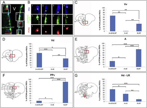
Quantification of Blbp and cyp19a1b-GFP (AroB) expression in proliferative radial glial cells in different neurogenic regions. A: Blbp (blue) and PCNA (red) staining on cyp19a1b-GFP (green) transgenic zebrafish line in the dorsomedian telencephalon. Proliferative cells (PCNA) can be Blbp-positive (inset 1), GFP-positive (inset 2), Blbp/GFP-positive (inset 3) or Blbp/GFP-negative (inset 4). The double negative group has been previously proposed to correspond to further committed precursors suggested to be neuroblasts ( März et al., 2010). B: Columns 1, 2, 3 and 4 corresponds to the respective insets in A for each channel. In 1, asterisks point to a proliferative cells displaying Blbp staining but not GFP. In 2, asterisks highlight a proliferative cells displaying GFP staining but not Blbp. In 3, asterisks show a proliferative cell displaying both GFP and Blbp staining, and neither staining in 4. C-G: In each region of interest, proliferative (PCNA) radial glial cells were checked for the expression of Blbp and cyp19a1b-GFP (AroB). Counting was performed on 6 adult female zebrafish (n = 6), on 325 cells for the Vv, 270 for the Vd, 205 for the A, 158 for the PPv, 185 for the Hc Div and 205 for the nucleus of the LR. The selected regions correspond to the zebrafish brain atlas ( Wullimann et al., 1996) section level number 71 for the Vv and Vd; 131 for the A; 168 for the PPv and the Hd LR. Statistical analysis was performed using one-way ANOVA. Error bars correspond to standard errors (n = 6 zebrafish). * 0,01 < p < 0,05; ** 0,01 < p < 0,0001; ***p < 0,0001. |
Reprinted from Gene expression patterns : GEP, 20(1), Diotel, N., Vaillant, C., Kah, O., Pellegrini, E., Mapping of Brain lipid binding protein (Blbp) in the brain of adult zebrafish, co-expression with aromatase B and links with proliferation, 42-54, Copyright (2016) with permission from Elsevier. Full text @ Gene Expr. Patterns