
Fig. 1
- ID
- ZDB-IMAGE-160304-10
- Publication
- Diotel et al., 2016 - Mapping of Brain lipid binding protein (Blbp) in the brain of adult zebrafish, co-expression with aromatase B and links with proliferation
- All Figures
- Figures for Diotel et al., 2016
|
Fig. 1
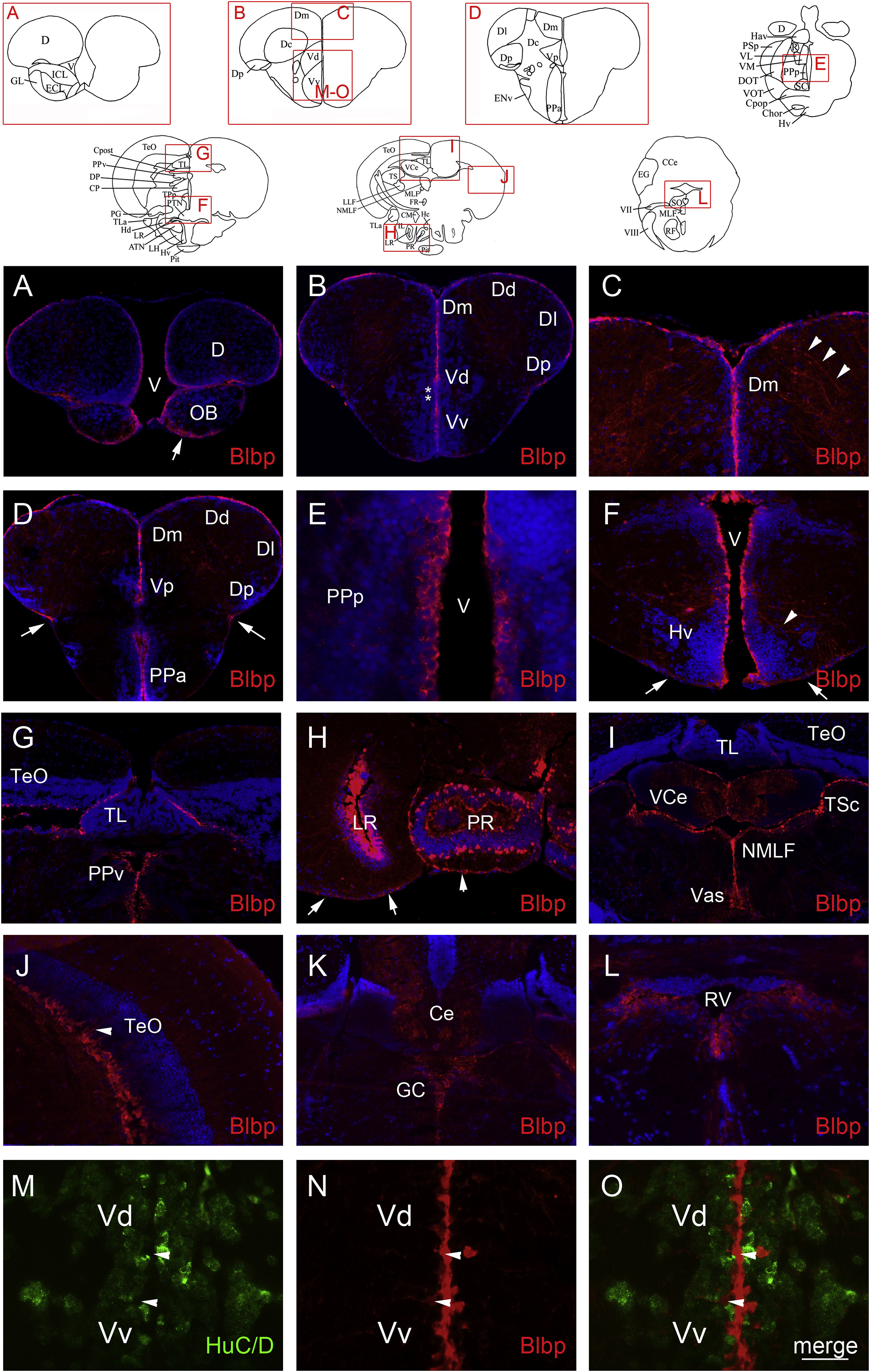
Blbp distribution in the brain of adult zebrafish. A-L: Blbp (red) immunohistochemistry on paraffin brain sections of adult zebrafish. DAPI counterstaining (blue) allows the visualization of cell nuclei. Blbp staining is detected in radial glial cells along the ventricular layers of the dorsal telencephalic area at the junction with the olfactory bulbs (A), in the ventral and dorsal nuclei of the ventral telencephalon, and in the dorsomedian, the dorsolateral, the dorsal and the posterior zone of the dorsal telencephalic area (B-D). In the RMS region (see asterisks in B), no Blbp staining was detected. Blbp-positive radial glial cells are also observed in the preoptic area and the hypothalamus (D-F). Strong Blbp staining is notably observed in cells surrounding the lateral and posterior recess of the hypothalamus (H). Radial glial cells from the periglomerular gray zone of the optic tectum are also positive for Blbp and extend long cytoplasmic process to the periphery of the brain (G and J, see arrowheads in J), in the periventricular pretectal nucleus (G). Blbp staining around the torus longitudinalis, the torus semicircularis, in the nucleus of the medial longitudinal fascicle (NMLF) and along the vascular lacuna of area postrema (Vas) correspond to Blbp-positive fibers (G and I). Few positive cells are observed in the valvula of the cerebellum and the cerebellum (I and K) and lining the rhombencephalic ventricle (K-L). Arrowheads highlight some long radial cytoplasmic processes crossing the brain parenchyma to reach the periphery of the brain (C, F and J). Arrows show the end of radial glial process at the pial surface (A, D, F and H). M-O: Blbp (red) and HuC/D (green, neuronal marker) double immunohistochemistry reveals that Blbp is not expressed in mature neurons. Arrowheads indicate Blbp positive radial glial cells that do not express the HuC/D neuronal marker. Bar: 16 µm (E, J, M, N and O); 50 µm (C, F, G and L); 100 µm (A, B, D, H, I and K).
Reprinted from Gene expression patterns : GEP, 20(1), Diotel, N., Vaillant, C., Kah, O., Pellegrini, E., Mapping of Brain lipid binding protein (Blbp) in the brain of adult zebrafish, co-expression with aromatase B and links with proliferation, 42-54, Copyright (2016) with permission from Elsevier. Full text @ Gene Expr. Patterns