- Title
-
ADSSL1 mutation relevant to autosomal recessive adolescent onset distal myopathy
- Authors
- Park, H.J., Hong, Y.B., Choi, Y.C., Lee, J., Kim, E.J., Lee, J.S., Mo, W.M., Ki, S.M., Kim, H.I., Kim, H.J., Hyun, Y.S., Hong, H.D., Nam, K., Jung, S.C., Kim, S.B., Kim, S.H., Kim, D.H., Oh, K.W., Kim, S.H., Yoo, J.H., Lee, J.E., Chung, K.W., Choi, B.O.
- Source
- Full text @ Ann. Neurol.
|
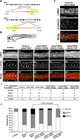
Loss-of-function of zebrafish adssl1 phenocopies distal myopathy. (A) Diagram of designed morpholinos (MOs) for zebrafish adssl1. Rectangles, black lines, and red bars indicate exons, introns, and location of MOs, respectively. (B) About 5 somites in the red box were counted to quantify the muscle phenotype. A = anterior; P = posterior. (C) Class II phenotype in the adssl1 MO2-injected zebrafish larva at 24 hours postfertilization (hpf). MHC = myosin heavy chain. (D–D′) Muscle morphology of uninjected control zebrafish larva at 24 hpf. (E–E′) Defects in the muscle of the zebrafish larva injected with adssl1 MO. (F–F′) The morphology in the muscle of the zebrafish larva coinjected with adssl1 MO and the mRNA of human ADSSL1 D304N. (G–G′) Disruption in the muscle of the zebrafish larva coinjected with adssl1 MO and the mRNA of human ADSSL1 I350fs. (H–H′) Rescued muscle phenotype by coinjection of human ADSSL1 (wild-type [WT]) mRNA and adssl1 MO in zebrafish larva at 24 hpf. (I) The table shows the embryo numbers with the muscle phenotype in each genotype. (J) A graph shows the quantified data. Asterisks indicate a disorganized fiber pattern (C) and loosely packed myofibers with occasional prominent gaps (E–G). Arrows point out the breakage of the myosepta. (C) Cells detached from the myosepta (E′–G′). Scale bars = 40 µm. PHENOTYPE:
|

