
- Title
-
Dynamic remodeling of the extra cellular matrix during zebrafish fin regeneration
- Authors
- Govindan, J., Iovine, M.K.
- Source
- Full text @ Gene Expr. Patterns
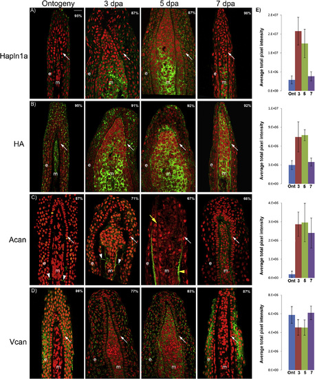
Immunostaining and histochemistry for Hapln1a-ECM components during the time course of regeneration. Longitudinal fin sections were treated with the respective primary antibodies and detected using the corresponding secondary antibody conjugated with Alexa Fluor– 488 (green). Propidium iodide (nuclei) is used as the counter stain (red). For each time point the percentage of sections showing similar expression pattern is denoted in each panel (n = 40–65 sections). (A) Immunostaining for Hapln1a; (B) histochemical detection of HA using biotin-HA binding protein; (C) immunostaining for Acan, the arrowhead identifies the joints and (D) immunostaining for Vcan. (E) The graph illustrates the overall change in the expression level during the time course of regeneration for each component. Efforts to compare expression levels between components were not completed. Arrows identify the basal layer of epithelium (BLE); yellow arrowhead identifies lepidotrichia and yellow arrow identifies actinotrichia; m, mesenchyme; e, epidermis; dpa, days post amputation. Scale bar is 20 µm. EXPRESSION / LABELING:
|
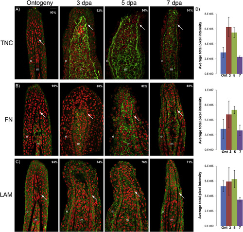
Immunostaining for TNC, FN and LAM during the time course of regeneration. Longitudinal fin sections were treated with the respective primary antibodies and detected using the corresponding secondary antibody conjugated with Alexa Fluor– 488 (green). Propidium iodide (nuclei) is used as the counter stain (red). For each time point under study the percentage of sections showing similar expression pattern is denoted in each panel (n = 40–65 sections). (A) Immunostaining for TNC; (B) immunostaining for FN and (C) immunostaining for LAM. (D) The graph illustrates the overall changes in the expression levels of each of the component under study during the time course of regeneration. Efforts to compare expression levels between components were not completed. Arrows identify the basal layer of epithelium (BLE); m, mesenchyme; e, epidermis dpa, days post amputation. Scale bar is 20 µm. EXPRESSION / LABELING:
|
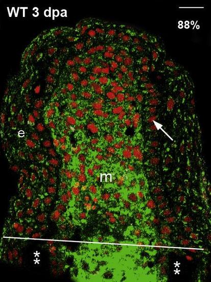
HA is upregulated in the stump of the regenerating fin. Wild type (WT), 3 dpa, longitudinal fin sections were treated biotin-HA binding protein and detected using the streptavidin conjugated with Alexa Fluor– 488 (green). Propidium iodide (nuclei) is used as the counter stain (red). White line represents the amputation plane and the asterix (**) marks the bone. The percentage of sections showing similar expression pattern is denoted in each panel (n = 40–65 sections). Arrow identifies the basal layer of epithelium (BLE); m, mesenchyme; e, epidermis; dpa, days post amputation. Scale bar is 20 µm. |
Reprinted from Gene expression patterns : GEP, 19(1-2), Govindan, J., Iovine, M.K., Dynamic remodeling of the extra cellular matrix during zebrafish fin regeneration, 21-9, Copyright (2015) with permission from Elsevier. Full text @ Gene Expr. Patterns