- Title
-
Whole-exome Sequencing Identifies a Variant in TMEM132E Causing Autosomal-Recessive Nonsyndromic Hearing Loss DFNB99
- Authors
- Li, J., Zhao, X., Xin, Q., Shan, S., Jiang, B., Jin, Y., Yuan, H., Dai, P., Xiao, R., Zhang, Q., Xiao, J., Shao, C., Gong, Y., Liu, Q.
- Source
- Full text @ Hum. Mutat.
|
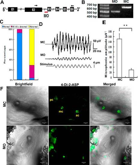
Tmem132e knockdown affects hair cell function in the otic cavity in zebrafish. A: Schematic representation of a partial pre-mRNA map showing the predicted outcome. The exons are in boxes, labeled with the exon number, and the introns are represented by sold lines. The location of MO is indicated with a red bar. Arrows show the locations of the primers used to amplify the splicing products in RT-PCR. B: The RT-PCR products amplified from MO and MC larvae confirm that the injected MO disrupts the normal splicing of tmem132e. C: Histogram of the swimming behavior of MO and MC larvae. D: Microphonic potentials in inner ear of 5-dpf larvae injected with control MC (top trace) and tmem132e MO (second trace). The bottom trace indicates 2-µm amplitude of stimulation. E: Average peak-to-peak amplitude of microphonic potential with MC and tmem132e MO. Number of larvae: MC, 10; MO, 15. Data are mean ± SEM. **P < 0.02. F: Micropressure injection of 4-Di-2-ASP (green) into the otic cavities labels hair cells in the anterior cristae (ac), medial cristae (mc), and posterior cristae (pc) of MC but not MO fish. Bars, 20 µm. PHENOTYPE:
|
|
Defective neuromast hair cells of L-PLL in zebrafish. A: 4-Di-2-ASP (green) labels neuromasts at 5 dpf. Exposure of a MC larva to 4-Di-2-ASP labels neuromasts (green). Labeling is absent in an MO larva. Coinjection of wild-type RNA coding for wild-type human TMEM132E mostly rescues a MO larva, restoring labeling in most neuromasts. Coinjection of MT RNA barely rescues a MO larva. MT, mutant. Bars, 200 µm. B: Histogram of neuromast numbers of both sides of L-PLL at 5 dpf. C: Scanning electron micrograph of a neuromast from the L-PLL. One midtail neuromast from the PLL of a MC larva at 5 dpf shows 12-long kinocilia emerging from short clusters of stereocilia and the pair of semilunar periderm cells surrounds each neuromast, which are highlighted with yellow dotted line in C (MC). In the MO larvae, the center of a neuromast displays fewer and shorter kinocilia and stereocilia. In addition, the semilunar periderm cells appear distorted. PHENOTYPE:
|


