
- Title
-
Histopathologic alterations associated with global gene expression due to chronic dietary TCDD exposure in juvenile zebrafish
- Authors
- Liu, Q., Spitsbergen, J.M., Cariou, R., Huang, C.Y., Jiang, N., Goetz, G., Hutz, R.J., Tonellato, P.J., Carvan, M.J.
- Source
- Full text @ PLoS One
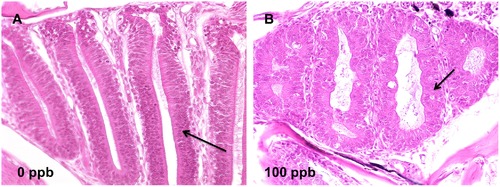
TCDD caused cystic degeneration in nasal epithelium in zebrafish after 42 d of dietary exposure. A) Well differentiated, orderly, uniform lamella (arrow) of zebrafish in 0 ppb group; B) cystic degeneration of nasal epithelium (arrows) was found in 100 ppb TCDD-treated zebrafish at 42 d (40X). |
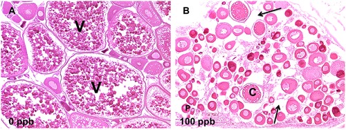
TCDD caused disruption of ovary development in zebrafish at 42 d. A) Ovulated vitellogenic follicles (V) in mature ovary of zebrafish in 0 ppb group; B) immature ovary in fish treated with 100 ppb TCDD. Ovary lacks vitellogenic follicles. Moderate interstitial edema (arrows) was observed; only previtellogenic oocytes are present: cortical alveolar oocytes (C) and perinucleolar oocytes (P) (10X). |
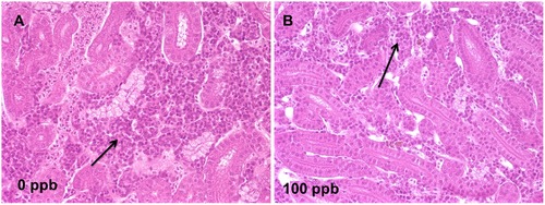
TCDD induced hemopoietic hypoplasia in kidney at 42 d. A) Normal hemopoietic tissue (arrow) in interstitium of anterior kidney of zebrafish in 0 ppb group; B) depletion of hemopoietic tissue (arrow) from interstitium of anterior kidney of zebrafish in 100 ppb group at 42 d (40X). |
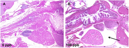
TCDD caused pericardial edema in zebrafish at 42 d. A) Normal heart in zebrafish of 0 ppb group; B) pericardial edema (arrow) in fish fed 100 ppb TCDD at 42 d (5X). |