
- Title
-
Soybean meal induces intestinal inflammation in zebrafish larvae
- Authors
- Hedrera, M.I., Galdames, J.A., Jimenez-Reyes, M.F., Reyes, A.E., Avendaño-Herrera, R., Romero, J., and Feijóo, C.G.
- Source
- Full text @ PLoS One
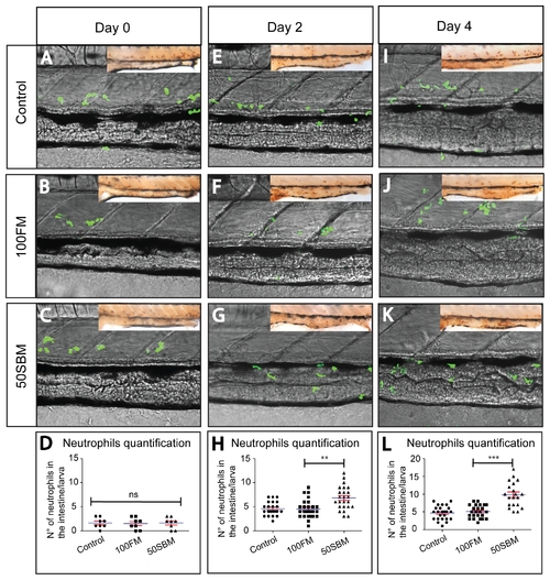
Soybean meal triggers neutrophil migration to the intestine after two days of feeding. Tg(mpx:GFP) transgenic larvae were analyzed in vivo by confocal microscopy to monitor neutrophil infiltration into the intestine at day 0 (A–C), 2 (E–G) and 4 (I–K) of feeding with control (pellet) and experimental diets (100FM diet and50SBM diet). (D, H, L) The number of intestine-infiltrated neutrophils was quantified by immunohistochemistry in the same larvae analyzed by confocal microscopy (inset). The results indicated that fish meal did not activate the immune response after 2 or 4 days of feeding. In the case of soybean meal, the number of infiltrated neutrophils was increased from day 2. Five larvae per condition in three different experiments were analyzed per time point by confocal microscopy. For immunohistochemistry analysis, at least 15 larvae per condition per time point in three different experiments were performed. Statistical analysis was conducted using one-way ANOVA. **p value < 0.01; ***pvalue < 0.001. EXPRESSION / LABELING:
|
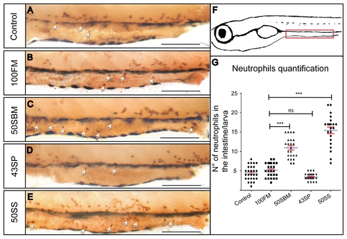
Soy saponin, but not soy protein, increases neutrophil migration to the intestine. Two compounds of soybean meal (50SBM diet) (C), soy protein (43SP diet) (D) and soy saponin (50SS diet) (E), were analyzed to determine their immunological effect. (G) An increase of neutrophils in the intestine (white arrow) was observed after 4 days of feeding in the soy saponin group (and soybean meal), but not in the soy protein group. (F) Scheme representing the intestine territory analyzed (red rectangle). At least 15 larvae per condition were analyzed in three different experiments. Statistical analysis was performed using one-way ANOVA. ns (no significance) *p value < 0.05; ***p value < 0.001. Scale bar = 200μm. EXPRESSION / LABELING:
|

Intestine morphology of a 9dpf larva. (A) Larva lateral view with the intestine delineated (dotted red line). Sagittal (B) and transversal (C–E) paraffin sections stained with H&E and alcian blue. The intestine is divided in 3 different segments: anterior (C) middle (D) and posterior (E). The arrow indicates goblet cells and the arrowhead indicates a supranuclear vesicle. Scale bar = 200μm in B; 50µm in C, D and E. |
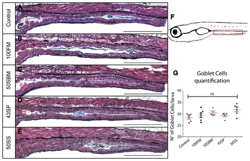
The number of goblet cells in larvae fed with the different diets does notchange after 4 days of treatment. Goblet cells quantification (F) in the intestine was performed in paraffin sections stained with H&E and alcian blue. No change in these mucus-secreting cells was observed between control groups (A) and experimental diets (B–E). At least six larvae were analyzed per condition in three different experiments. Statistical analysis was performed using one-way ANOVA. ns (no significance). Scale bar = 200μm. |
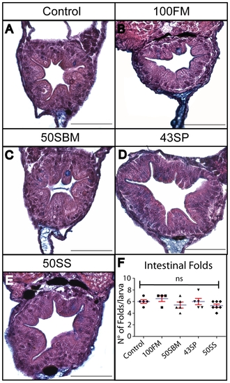
Intestinal folds did not disappear in larvae fed with Soybean meal or soy saponin diets. Transversal section of the mid-intestine, stained with H&E and alcian blue, of larvae fed with the different diets analyzed. The quantification of intestinal folds number in each case indicated that there is no difference between control and experimental diets (F). At least six larvae were analyzed per condition in three different experiments. Statistical analysis was performed using one-way ANOVA. ns (no significance). Scale bar = 50μm. |
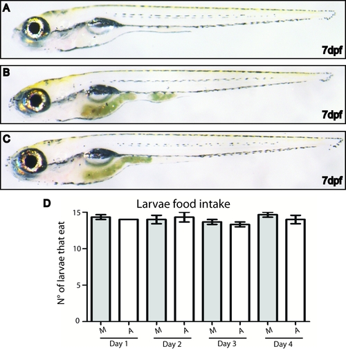
Figure S2. Monitoring larvae food ingestion. Lateral view of a 7dpf larva before feeding (A) and after food ingestion (B, C). Food ingestion can be easily verified by observing food presence in the gut. (D) Quantification of the number of larvae that eat in every feeding. |