
- Title
-
Co-Expression of VAL- and TMT-Opsins Uncovers Ancient Photosensory Interneurons and Motorneurons in the Vertebrate Brain
- Authors
- Fischer, R.M., Fontinha, B.M., Kirchmaier, S., Steger, J., Bloch, S., Inoue, D., Panda, S., Rumpel, S., and Tessmar-Raible, K.
- Source
- Full text @ PLoS Biol.
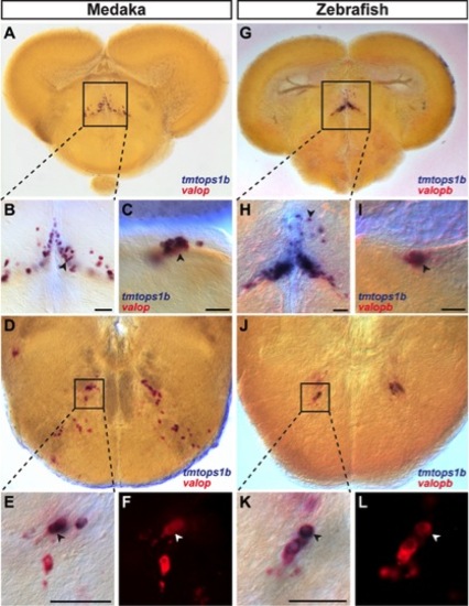
Two-color ISH of tmt-opsin1b (blue) and val-opsins (red/red fluorescence) on coronal medaka (A–F) and zebrafish (G–L) sections. Magnifications are indicated as boxes. Scale bars, 50 μm. Tmtops1b and valopb co-expression in the central posterior thalamic nucleus of medaka (A–B) and zebrafish (G–H), in the dorsal tegmental nucleus in medaka (C) and zebrafish (I) and in the facial nerve nucleus in medaka (D–F) and zebrafish (J–L). EXPRESSION / LABELING:
|
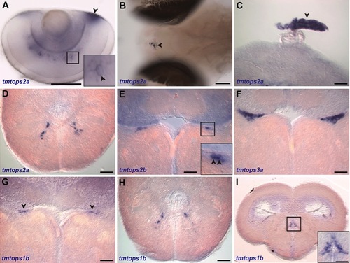
Tmt-opsin expression in zebrafish reveals evolutionary conservation of expression domains. ISH on 6 dpf zebrafish larvae (A, B) and coronal sections of the adult brain (C–I). Scale bars, 100 μm. (A) tmtops2a expression in amacrine cells and the annular ligament (black arrowhead). Pineal expression of tmtops2a in larval (B) and adult brains (C). tmtops2a in the facial nerve nucleus (D) and the dorsal tegmental nucleus (E). The dorsal tegmental nucleus was also stained for tmtops3a (F) and tmtops1b (G). tmtops1b is also present in the facial nerve nucleus (H) and the central posterior thalamic nucleus (I). |