
- Title
-
An artificial promoter construct for heat-inducible misexpression during fish embryogenesis
- Authors
- Bajoghli, B., Aghaallaei, N., Heimbucher, T., and Czerny, T.
- Source
- Full text @ Dev. Biol.
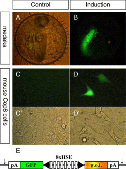
Activation of the HSE promoter in medaka embryos and in cell culture. The gfp:HSE:luc DNA construct was co-injected with meganuclease into one-cell stage medaka embryos (A and B) or transfected in mouse Cop8 cells (C, C2, D, and D2). The injected embryos were divided into a control group and a test group. Embryos of the untreated control group (A) were gfp-negative. Embryos of the test group were treated at 39°C for 2 h, resulting in strong gfp expression (B). Typical embryos (stage 24) are shown for both groups. After transfection into mouse Cop8 cells, control plates remained gfp-negative (C). Treatment at 44°C for 2 h induced a strong gfp response in the transfected cells (D). (C2 and D2) are the corresponding brightfield views for C and D, respectively. (A, B, C, and D) are fluorescent images, background light was added for image (A) to visualize the otherwise gfp-negative embryo. A schematic presentation of the HSE promoter is depicted in (E). The artificial promoter contains eight multimerized heat shock elements flanked by two minimal promoters in opposed orientation (black arrowhead). Gfp on one side and the gene of interest (luciferase or Fgf8) at the other side are expressed from the bicistronic promoter. The vector is flanked by I-SceI meganuclease sites (arrows). Abbreviations: od, oil droplet; pA, SV40 polyadenylation signal; HSE, heat shock element; g.o.i., gene of interest. |
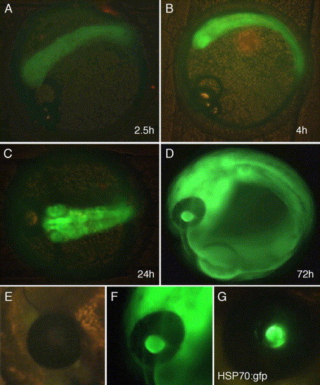
Stable integration of a HSE construct into the medaka genome. (A–D) show a time course of gfp expression in a gfp:HSE:luc transgenic line. After heat treatment at 39°C (2 h), gfp expression was first detected after 2.5 h (A). Increasing signal intensity was seen after 4 h (B) and 24 h (C). Gfp activity remained for several days (D, 72 h). Note the reduction of gfp signal in the eye, but not the lens, due to pigmentation. Lenses of uninduced transgenic embryos were background-free also in the lens (E), heat treatment induced a strong response in all tissues, including the lens (F). Wild-type embryos injected with HSP70:gfp developed strong gfp activity in the lens without induction (G). |
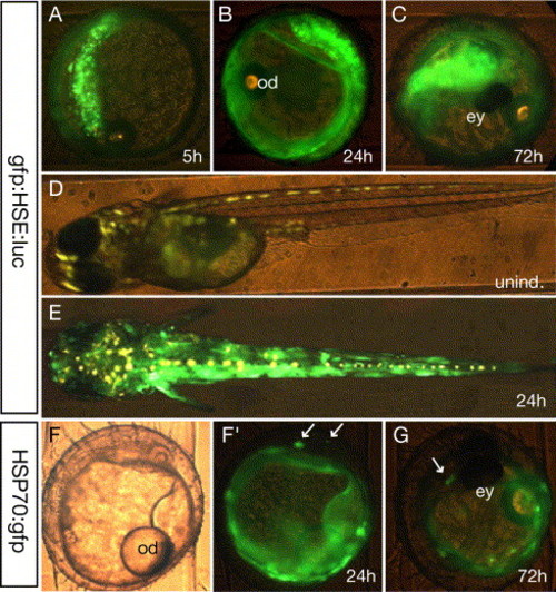
Transient misexpression with heat stress-inducible constructs. Medaka embryos were co-injected at the one-cell stage with gfp:HSE:luc (10 ng/µl) and meganuclease enzyme. Heat treatment was applied after 24 h at embryonic stage 19 (39°C/2 h). Gfp expression was recorded 5 h (A), 24 h (B), and 72 h (C) after induction. Uninduced embryos were grown until hatching and did not show any gfp expression (D). Yellow staining originates from autofluorescing cells. These larvae were induced (39°C/1 h) and exhibited a strong response after 24 h (E). For comparison, embryos were injected the same way with the HSP70:gfp construct and induced under the same conditions (F, F2, and G). Gfp signals were preferentially observed in yolk cells (F2), gfp-positive cells within the embryo are marked by arrows. (G) The same embryo 48 h later. F is a brightfield view of F2. Abbreviations: od, oil droplet; ey, eye. |
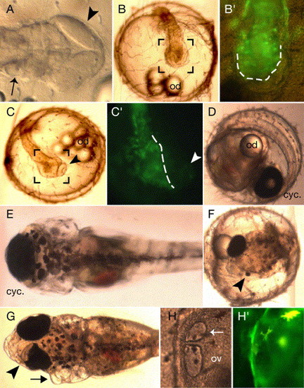
Phenotypes of medaka embryos misexpressing Fgf8. Injection of Fgf8 mRNA and CMV:Fgf8 results predominantly in a strong eye/forebrain phenotype (A). In most cases, the forebrain is dramatically reduced, but more posterior structures appear normal (otic vesicle marked by an arrow). The midbrain appears dramatically expanded (arrowhead) and structures anterior to the midbrain became lost. (B and C) show examples of observed eye phenotypes after induction of the gfp:HSE:Fgf8 DNA at stage 14. (B2 and C2) gfp expression in the area encompassing the black square marks in the corresponding images (B and C). Embryos developing cyclopic eyes (B) exhibit misexpression not within the eye, but in the adjacent tissue (B2 dotted line indicates the border between the eye and the forebrain). Loss of one eye (C) correlated with misexpression on the same side of the embryo (C2 the dotted line demarcates gfp-positive and -negative cells), the other eye is marked by arrowheads. D and E show the embryo with the cyclopia phenotype (B) at a later embryonic stage (D) and a larval stage (E). An embryo induced at the two-somite stage (stage 19) exhibited reduced size of one eye marked by an arrowhead (F). A pigmentation phenotype in one eye (arrowhead) and an expanded otic vesicle (arrow) were seen for a larva induced during mid-gastrula (G). Ectopic otic vesicles were repeatedly observed, a magnification of such an embryo is shown in (H); the ectopic otic vesicle is marked by an arrow. Note that gfp, marking the Fgf8 misexpressing cells, is not seen within in the vesicle (H2); autofluorescing medaka cells are marked by an arrowhead. Abbreviations: cyc., cyclopic eye; od, oil droplet. |
Reprinted from Developmental Biology, 271(2), Bajoghli, B., Aghaallaei, N., Heimbucher, T., and Czerny, T., An artificial promoter construct for heat-inducible misexpression during fish embryogenesis, 416-430, Copyright (2004) with permission from Elsevier. Full text @ Dev. Biol.