- Title
-
A cross-species analysis of Satb2 expression suggests deep conservation across vertebrate lineages
- Authors
- Sheehan-Rooney, K., Pálinkášová, B., Eberhart, J.K., and Dixon, M.J.
- Source
- Full text @ Dev. Dyn.
|
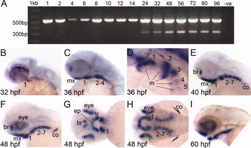
Expression of zebrafish satb2 is conserved compared with other vertebrates. A: A duplex reverse transcriptase-polymerase chain reaction (RT-PCR) designed to simultaneously amplify β-actin (555 bp) and satb2 (306 bp) indicated that satb2 was expressed from 24 to 96 hours postfertilization (hpf). The negative control (-ve) in which the cDNA is replaced with sterile water did not produce an amplicon. B–I: satb2 expression was analyzed using the satb2 antisense riboprobe. Images B, C, E, F, and I are lateral images with anterior facing left. Image D is a higher magnification, dorsolateral view of the pharyngeal arches in C. Image G is a frontal view of the embryonic face, and image H is a dorsal view of the embryo shown in F and G. B: satb2 expression was first detected at 32 hpf in the most anterior–ventral aspect of the first pharyngeal arch/mandible. C,D: By 36 hpf, satb2 expression was detected in the ventral mesenchyme of pharyngeal arches 1–5 and maxillary condensations. D: Expression in the pharyngeal arches clearly marked the mesenchyme surrounding a core of satb2-negative mesoderm. E,F: This expression pattern persisted but became more intense such that by 40 hpf satb2 expression was observed in all the pharyngeal arches, forebrain, scapulocoracoid of the pectoral fin and, by 48 hpf, the eye. G: Frontal views of 48 hpf larvae showed satb2 expression marking the trabeculae and ethmoid plate of the anterior neurocranium. H: Expression in the arches appeared to mark areas that form the cartilage elements of the pharyngeal skeleton. I: The expression pattern of satb2 continued until 60 hpf and thereafter declined. mx, maxillary condensations; 1–7, pharyngeal arches 1–7; m, mesoderm; br, forebrain; co, scapulocoracoid; tr, trabeculae; ep, ethmoid plate. |
|
Zebrafish satb2 expression marks the prechondrogenic mesenchyme. A–J: Transverse (A–C,F–J) and parasagittal (D,E) sections are shown. Wild-type zebrafish embyos at 38 hours postfertilization (hpf; A–E) and 48 hpf (F–J) were sectioned following whole-mount in situ hybridization using the zebrafish antisense satb2 riboprobe. A–C: Progressive sections along the anterior–posterior axis of the embryo at 38 hpf showed satb2 expression in the brain and the mesenchymal precursors of the ethmoid plate and trabeculae while the ectoderm was satb2-negative. D,E: Parasagittal sections showed abundant satb2 expression in the mesenchyme of the maxillary condensations and pharyngeal arches 1–5 (1–5) that surrounded a core of satb2-negative mesoderm. F–J: Progressive sections along the anterior–posterior axis of the embryo at 48 hpf showed satb2 expression in the brain and eyes. G,H: satb2 expression was subsequently detected in the prechondrogenic mesenchyme of the ethmoid plate and trabeculae but not in the overlying ectoderm. I,J: Further posteriorly, the prechondrogenic mesenchyme of all seven pharyngeal arches was satb2 positive as well as scapulocoracoid of the pectoral fin. br, brain; ep, ethmoid plate; tr, trabeculae; e, ectoderm; mx, maxillary condensations; 1–5, pharyngeal arches 1–5; m, mesoderm; co, scapulocoracoid; oc, oral cavity; pf, pectoral fin. |
|
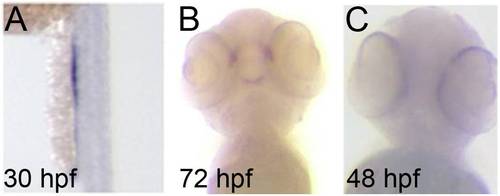
Developmental expression profile of satb2 in zebrafish. A: Whole-mount in situ hybridization using the antisense satb2 riboprobe revealed expression in the pronephric duct at 30 hours postfertilization (hpf). B: satb2 was expressed until 72 hpf, but which time expression was limited to the mouth. C: satb2 expression was not observed in the negative control in which the sense satb2 riboprobe was used. EXPRESSION / LABELING:
|

Unillustrated author statements |


