- Title
-
Alternative splicing of sept9a and sept9b in zebrafish produces multiple mRNA transcripts expressed throughout development
- Authors
- Landsverk, M.L., Weiser, D.C., Hannibal, M.C., and Kimelman, D.
- Source
- Full text @ PLoS One
|
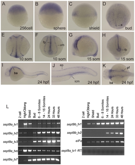
Expression of sept9 genes during zebrafish development. Detection of sept9 mRNA was carried out by whole-mount in situ hybridization using a probe targeted to all sept9 isoforms on staged embryos from 256 cells to 24 hpf. Images in A–C are lateral views, animal pole to top; D and E are dorsal views, anterior to top; F and H are dorsal posterior views; G, I and J are lateral views, K is a dorsal view, anterior to left. A–C: sept9 transcripts are ubiquitously expressed at early developmental states. D: At bud stage, sept9 is expressed in endoderm and axis. E–H: sept9 is expressed in the floor plate, ventral mesoderm and tail bud during segmentation. I–K: At 24 hpf, sept9 is expressed throughout the epidermis, branchial arches, pectoral fin, and in the intermediate cell mass. L: Transcript specific primers were used to detect sept9a and sept9b transcripts in various stages of development by RT-PCR. sept9a_tv 2, 3, and α are expressed maternally. Amplification of eIFα and total RNA without addition of reverse transcriptase were used as controls. a, axis; ep, epidermis; fp, floorplate; icm, intermediate cell mass; tb, tail bud; vm, ventral mesoderm. |
|
Characterization of sept9a morphant and overexpression embryos. Embryos were injected at the one-cell stage with morpholinos targeted to all sept9a transcripts (MO5), sept9a_tv1 only (MO2), mismatch controls (MO5MM, MO2MM), or sept9a_tv1 mRNA with and without MO2. Morphants shown were injected with 2.5 ng morpholino. At 48 hpf, the phenotypes were assessed by morphological criteria, according to severity. A,D: Class I morphants had defects in epidermal integrity and yolk extension and minor curvature of the tail. B,E: Class II morphants had a curved body axis in addition to the defects observed in class I. Class III morphants had a severely shortened body axis (data not shown). Arrows indicate yolk extension defects. All classes exhibited defects in blood circulation. G: Coinjection of 1 pg of sept9a_tv1 mRNA with 5 ng of MO2 partially rescued the observed phenotypes. H,I: Embryos injected with as little as 4 pg of sept9a_tv1 mRNA often had phenotypes similar to those of sept9a morphants including epidermal aggregates (arrow), blood pooling, and tail edema (bracket). (OE) indicates over expression. C,F: Control mismatch morpholinos did not present a phenotype. J: Graphical representation of MO classes at various concentrations. The number of embryos tested in each experiments is indicated by (n) on top of each column. K: sept9a splice morpholinos inhibit sept9a transcript splicing. RT-PCR analysis was performed on 24 hpf wild-type embryos, embryos injected with 2.5 ng MO5 or MO2 (pooled classes I and II), or 5 bp mismatch controls. MO5 embryos show a complete loss of sept9a_tv1 and sept9a_tv7. The presence of a low level of sept9a exons 3–5 transcripts in MO5-injected embryos may be due to maternal mRNA. MO2 embryos show a decrease in sept9a_tv1 compared to wild-type while the other transcripts are not affected. PHENOTYPE:
|
|
Knockdown and overexpression (OE) of sept9a_tv1 results in an increase in apoptotic cells in the tail. Embryos at the one-cell stage were injected with 2.5 ng of MO2 or 4 pg of sept9a_tv1 mRNA and analyzed for acridine orange (AO) staining at 24 hpf. A–C: The tail fin of wild-type embryos is negative for AO indicating few apoptotic cells. D–I: Both class II MO2 and sept9a_v1 OE embryos show an increase in AO staining indicating increased cell death. PHENOTYPE:
|

Unillustrated author statements |


