- Title
-
Expression of the zebrafish intermediate neurofilament Nestin in the developing nervous system and in neural proliferation zones at postembryonic stages
- Authors
- Mahler, J., and Driever, W.
- Source
- Full text @ BMC Dev. Biol.
|
nestin expression in zebrafish development. (A-D): nestin expression pattern during gastrula and early somitogenesis stages, (A) nestin is not expressed at 60% epibloy stage, (B) sense control. (C, D): First expression is detectable at 3 somite stage. (E, F-F″): Flat mount and cross sections of a 3 somite embryo. (G, sense control in H): Lateral view of 24 hpf embryo, nestin is expressed widely in CNS. (I, I′): 2 dpf, lateral view (I), dorsal view (I′) nestin is expressed in head ganglia and fin buds; insert in (I′) shows higher magnification of a fin bud; (I″) higher magnification of the spinal cord. nestin expression in the ventral root may correlate with glia or neuroblasts. (J-J″): At 3 dpf nestin expression becomes more restricted to proliferation zones, (J) lateral view, (J′, J″) higher magnifications, dorsal (J′) and lateral (J″) view. (K-K″): 4 dpf: nestin expression in the CNS is almost completely restricted to proliferation zones. Further expression is detected in the cranial ganglia, the gut and the craniofacial mesenchyme (K″). Abbreviations: ALLG: anterior lateral line ganglion; cm: craniofacial mesenchyme; FB: fin bud; g: gut; OG: octaval ganglion; no: notochord; SC: spinal cord; VG: vagal ganglion; VMP: ventral motoneuron precursors. (A-E, G-K″): anterior left, animal pole up, (F-F″): cross sections, dorsal up. Scale bars: 100 μm, except K: scale bar: 200 μm. |
|
In the developing CNS nestin expression becomes progressively restricted to proliferative zones. Analysis of nestin expression in the head by whole mount in situ hybridization. (A-C): lateral views, anterior left, dorsal up, eyes were removed for better visualization of the expression pattern in the DC. A mid-saggital focal plane is shown (D-O, except L): dorsal views, anterior left, the dorsoventral level of the focal plane is indicated at left. (L): lateral view. Abbreviations: CCe: Cerebellum; CMZ: ciliary marginal zone; DC: diencephalon; GCL: ganglion cell layer; MHB: midbrain hindbrain boundary; Pr: pretectum; Ret: retina; S: subpallium: TC: telencephalon; TeO: optic tectum. Scale bars: 100 μm. |
|
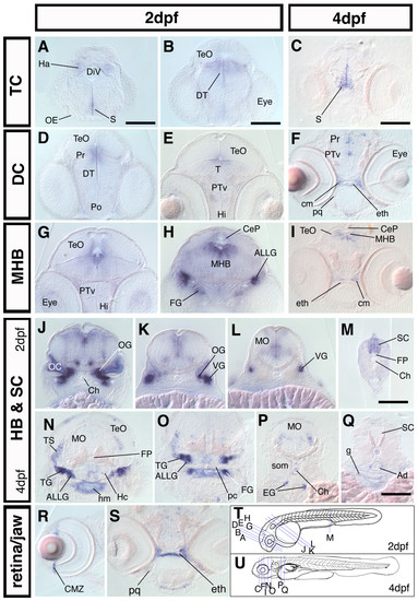
nestin is expressed in proliferative zones during the development of the CNS. Cross sections of 2 dpf and 4 dpf embryos; dorsal up. (A-S): frontal and transversal cross section of nestin expression along the rostrocaudal axis of the brain and in the spinal cord reveals expression in major proliferative zones The relative rostrocaudal levels of each section are indicated in T and U. (A-I): sections at levels of forebrain and midbrain as well as midbrain-hindbrain boundary. (J-Q): sections at the level of HB and SC, showing nestin expression in the cranial ganglia and the HB, MO, SC. (R): cross section through the eye of a 4 dpf embryo, nestin is expressed in the CMZ; (S): cross section of a 4 dpf embryo at the level of the ethmoid plate, nestin is expressed in the craniofacial mesenchyme adjacent to developing cartilage tissue. (T, U): scheme of a 2 dpf (T) and a 4 dpf (U) zebrafish embryo, the levels of the cross sections shown in A-S are indicated by blue lines. Abbreviations: Ad: Aorta dorsalis; ALLG: anterior lateral line ganglion; CeP: cerebellar plate; Ch: chorda dorsalis; cm: craniofacial mesenchyme; CMZ: ciliary marginal zone; DiV: diencephalic ventricle; DT: dorsal thalamus; eg: enteric ganglia; eth: ethmoid plate; FG: facial ganglion; FP: Floor plate; g: gut; Ha: habenula; Hc: caudal hypothalamus; Hi: intermediate hypothalamus; hm: head mesenchyme; MHB: midbrain hindbrain boundary; MO: medulla oblongata; OC: otic capsule; OE: olfactory epithelium; OG: octaval ganglion; pc: parachordal cartilage; pq: palatoquadrate; Po: preoptic region; Pr: pretectum; PTv: ventral part of the posterior tuberculum; S: subpallium; SC: spinal cord; som: somites; T: midbrain tegmentum; TeO: optic tectum; TG: trigeminal ganglion; TS: torus semicircularis; VG: vagal ganglion. Scale bars: 100 μm. EXPRESSION / LABELING:
|
|
Co-expression of nestin and pcna in the zebrafish forebrain. Double fluorescent ISH was performed for 2 dpf (A-I) and 3 dpf (J-L) zebrafish embryos. nestin is shown in green (A, D, G, J); pcna in red (B, E, H, K); overlay (C, F, I, L). Inserted pictures in (J-L) show a higher magnification of the boxed areas. (A-C): Overview over the proliferation zones in the 2 dpf zebrafish forebrain. (D-F): Higher magnification of the ventricular zone of the TC shows co-expression. (G-H): Higher magnification of the subventricular zone in the ventral DC reveals co-expression of nestin and pcna. (J-L): At 3 dpf cells co-expressing nestin and pcna are detectable along the walls of the telencephalic ventricle. Abbreviations: DC: diencephalon; Ret: retina; TC: telencephalon. Scale bar: 50 μm. EXPRESSION / LABELING:
|
|
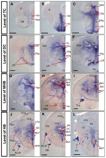
nestin expression in the juvenile 28 dpf old zebrafish brain. Analysis of nestin expression in the brain of the 28 dpf zebrafish by whole mount in situ hybridization using nestin anti sense mRNA probes on whole dissected brain. (A-L): selected 50 μm cross sections from a serially sectioned brain, the rostrocaudal levels of the sections are indicated at left; dorsal up. Pictures show only the left half of the brain. Dashed lines in J, K, L indicate the tracts of the cranial nerves. Abbreviations: ALLN: anterior lateral line nerve; CCe: corpus cerebelli; Cven: commissura ventralis rhombencephali; D: dorsal telencephalic area; DiV: diencephalic ventricle; EG: eminentia granularis; GC: griseum centrale; HaV: ventral habenular nucleus; Hc, Hd, Hv: caudal, dorsal, ventral zone of the periventricular hypothalamus; LC: locus coeruleus; LCa: locus caudalis cerebelli; LR: lateral recess of the DiV; MLF: medial longitudinal fascicle; NIII: oculomotor nucleus; NIV: trochlear nucleus; NMLF: nucleus of MLF; NLV: nucleus lateralis valvulae; OT: optic tract; PGZ: periventricular gray zone of the optic tectum; PPa, PPp: parvocellular preoptic nucleus, anterior, posterior part; PPv: periventricular pretectal nucleus, ventral part; PTN: posterior tuberal nucleus; RV: rhombencephalic ventricle; TeO: optic tectum; TeV: tectal ventricle; TelV: telencephalic ventricle; TL: Torus longitudinalis; TLa: torus lateralis; TN: thalamic nuclei; Val, Vam: lateral, medial division of valvula cerebelli; Vd, Vv: dorsal, ventral nucleus of ventral telencephalic area; TPp: periventricular nucleus of the posterior tuberculum; III: oculomotor nerve; VII: facial nerve; VIII: octaval nerve; Scale bars: 100 μm. EXPRESSION / LABELING:
|

Unillustrated author statements EXPRESSION / LABELING:
|


