
- Title
-
Deconstructing evolution of adult phenotypes: genetic analyses of kit reveal homology and evolutionary novelty during adult pigment pattern development of Danio fishes
- Authors
- Mills, M.G., Nuckels, R.J., and Parichy, D.M.
- Source
- Full text @ Development
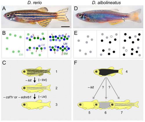
Danio adult pigment patterns, pigment pattern metamorphosis and genetic analyses, either real or envisaged. (A-C) Danio rerio. (A) Wild-type D. rerio exhibit around five melanophore stripes with light interstripes. (B) Early (left) to late (right) stages of adult pigment pattern metamorphosis. EM melanophores (light green) differentiate in a dispersed pattern then migrate (red tracks) toward developing stripes. LM melanophores (light blue) differentiate already at sites of stripe formation. Fully differentiated melanophores within adult stripes come from either EM (dark green) or LM (dark blue) melanophore populations. Other pigment cell classes are omitted for clarity. (C) EM melanophores are kit-dependent, whereas LM melanophores are kit-independent. Wild-type D. rerio (fish 1) with a full complement of melanophores and stripes (black). kit mutant D. rerio (fish 2) lacks EM melanophores but develops residual LM melanophores in stripes (gray) having fewer than wild-type numbers of melanophores. Fish doubly mutant for kit and csf1r or kit and ednrb1 (fish 3) lack body melanophores, as both EM and LM melanophores are missing. (D-F) Danio albolineatus. (D) Wild-type D. albolineatus have melanophores that are nearly uniformly dispersed, although a narrow interstripe is found posteriorly (arrow). (E). Pigment pattern metamorphosis involves the differentiation of initally dispersed melanophores (gray) that show little migratory behavior. Some of these melanophores differentiate fully (black) but many more die (open cells) than in D. rerio (Quigley et al., 2005). (F) Wild-type D. albolineatus (fish 4) with uniformly distributed melanophores (black), and possible phenotypes of kit mutant D. albolineatus. If D. albolineatus have only kit-dependent EM melanophores, a kit mutant should lack melanophores completely (fish 5). If distinct kit-dependent EM and kit-independent LM melanophores are present, however, a kit mutant should develop residual melanophores (fishes 6 or 7) that are fewer than in wild-type (gray uniform pattern or gray stripes). The pattern of these cells would reveal how different populations have contributed to the difference between wild-type D. rerio and wild-type D. albolineatus: if changes only in LM melanophores have occurred, then a kit mutant (lacking EM melanophores) might resemble wild-type D. albolineatus (fish 6); or, if changes only in EM melanophores have occurred, then a kit mutant D. albolineatus (fish 7) should resemble kit mutant D. rerio (fish 3). Fish ages: ∼6 months. Scale bar: in A, 4 mm for A,D. |

Isolation and molecular characterization of a kit mutant D. albolineatus and comparison with D. rerio. (A-D) Melanophore phenotypes of embryos at 60 hpf. (A) Wild-type D. rerio. (B) Wild-type D. albolineatus have fewer melanophores overall compared with D. rerio. (C) Homozygous kitb5 mutant D. rerio exhibit fewer melanophores, especially at sites distant from their origin in the neural crest (e.g. covering the yolk, arrow). An ectopic patch of melanophores (arrowhead) occurs posterior to the otocyst. (D) Homozygous wp.a14e1 D. albolineatus have fewer peripheral melanophores (arrow) and ectopic post-otic melanophores (arrowhead), although total melanophore numbers are greatly diminished compared with kit mutant D. rerio. (E) RT-PCR for amplicons (A1-A3) from D. albolineatus kit cDNAs. A1, lower transcript abundance in wp.a14e1 compared with wild type. A2, reveals the predicted 652 bp product in wild type but a 296 bp product in wp.a14e1. A3, failure to amplify a 234 bp product demonstrates absence of wild-type kit transcript in wp.a14e1. β-actin, loading control. (F) Schematics of D. albolineatus kit cDNAs and locations of amplicons A1-A3. Green, predicted signal sequence and transmembrane domain. Red, predicted kinase domains. In kitwp.a14e1 two exons are deleted, resulting in a frameshift with novel amino acids (orange) and a premature stop codon. Sequence analyses confirm that D. albolineatus kit is orthologous to D. rerio kit (kita), rather than a second kit locus identified in D. rerio, kitb (see Discussion) (Mellgren and Johnson, 2005). (G) Genomic structure of kit in wild type and wp.a14e1 mutant D. albolineatus. Shown are exons 4-6 and intervening introns (i4-i6). The region deleted in wp.a14e1 is shown in brown, and a novel inserted sequence is shown in orange. GenBank accession number for D. albolineatus kit: EF035010. PHENOTYPE:
|
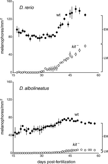
Quantitative analyses of melanophore accumulation in wild-type D. rerio and D. albolineatus and their respective kit mutants. In D. rerio (upper plot) wild-type melanophore densities (means±1 s.e.) are relatively constant during the first half of pigment pattern metamorphosis (∼15-36 dpf) but then increase dramatically during the second half of pigment pattern metamorphosis (∼37-49 dpf), corresponding to the appearance of new melanophores within stripes. Note that total melanophore numbers (as opposed to densities) increase throughout metamorphosis. In kit mutants there are no melanophores until late metamorphosis when residual LM melanophores develop. In D. albolineatus (lower plot), wild-type melanophore densities increase more slowly through metamorphosis and never reach the same maximum as in wild-type D. rerio. kit mutant D. albolineatus completely lack melanophores during early pigment pattern metamorphosis, then recover a few residual LM melanophores during late pigment pattern metamorphosis. Vertical bars to the right of plots indicate the inferred final contributions of EM and LM melanophores to the different pigment patterns. The timing of pigment pattern metamorphosis was somewhat delayed in these analyses compared with previous studies, presumably owing to slight differences in rearing conditions. PHENOTYPE:
|
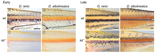
Pigment patterns at early metamorphosis and after late metamorphosis. (A) Wild-type D. rerio with initially dispersed metamorphic melanophores as well as nascent stripes bounding the primary interstripe (arrow). (B) Wild-type D. albolineatus has dispersed melanophores, which are largely absent from a corresponding interstripe region (arrow). (C,D) kit mutants of both species lack melanophores at early metamorphosis. (E) Wild-type D. rerio with a juvenile pigment pattern of two primary stripes bounding the primary interstripe and a third stripe developing further ventrally. (F) Wild-type D. albolineatus with melanophores relatively uniformly dispersed except for the interstripe region. Melanophores adjacent to the interstripe tend to be larger and darker. White bar indicates darkness owing to internal structures (aorta, vertebrae, etc.) rather than melanophores. (G) kit mutant D. rerio develop residual LM melanophores in stripes (large arrowheads) dorsal and ventral to the interstripe. (H) kit mutant D. albolineatus develop residual LM melanophores primarily in a stripe dorsal to the interstripe (large arrowheads), although a few melanophores are also found ventral to the interstripe (small arrowheads). Ages, A and C, 31 dpf; B and D, 28 dpf; E and G, 53 dpf; F and H, 60 dpf. Scale bars: in A, 500 μm for A-D; in E, 500 μm for E,G; in F, 500 μm for F,H. PHENOTYPE:
|
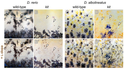
kit is required during a late step in melanophore development. Dorsal regions of late larval fish are shown before (A-D) and after (A'-D') incubation with L-dopa. Arrowheads, sites of melanoblasts before and after L-dopa treatment. (A,A') In wild-type D. rerio, previously melanized melanophores are present within the dorsal melanophore stripe and on dermal scales (arrow). Very few L-dopa+ melanoblasts are revealed (arrowheads). (B,B') kit mutant D. rerio exhibit more undifferentiated melanoblasts than wildtype D. rerio. (C,C') In wild-type D. albolineatus, many more L-dopa+ melanoblasts are present than in wild-type D. rerio. (D,D') kit mutant D. albolineatus have similar numbers of L-dopa+ melanoblasts to wild-type D. albolineatus. PHENOTYPE:
|

kit-dependent and kit-independent regenerative melanophores in D. albolineatus. (A) In wild-type D. albolineatus, many regenerative melanophores (arrowheads) develop by 10 dpa and ultimately form a pattern indistinguishable from that of the unamputated fin. These cells differentiate de novo and have not migrated in from unamputated regions, as revealed by initially rearing fish in the melanin synthesis inhibitor phenylthiourea, allowing new and previously differentiated melanophores to be distinguished from one another (data not shown) (see Rawls and Johnson, 2000). Vertical bars, amputation planes. Arrows, melanophores already present in the unamputated fin. (B) In kit mutant D. albolineatus, regenerative melanophores do not develop by 10 dpa. Dark regions along edges of lepidotrichia are shadows (e.g. black arrow), not melanophores. (C) After longer periods (≥20 dpa) a few kit-independent regenerative melanophores differentiate (arrowhead), although fins never recover melanophore numbers equivalent to wild-type regenerates. Scale bar: in C, 500 μm for A-C. |