
Image
|
Figure Caption
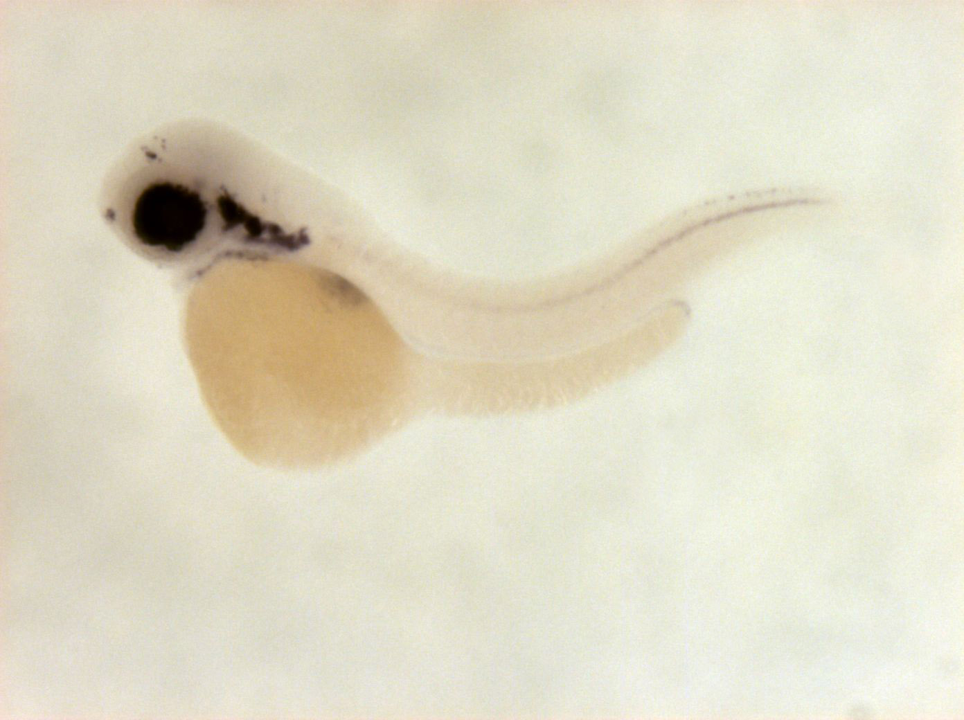
Fig. 6 cells in retina, one endoderm primordium, cranial ganglia (stronger in trigeminal), hindbrain, spinal chord neurons, branchial arches, cells in pretectum and telencephalon
Orientation
| Preparation | Image Form | View | Direction |
| whole-mount | still | side view | anterior to left |
Figure Data