
Image
|
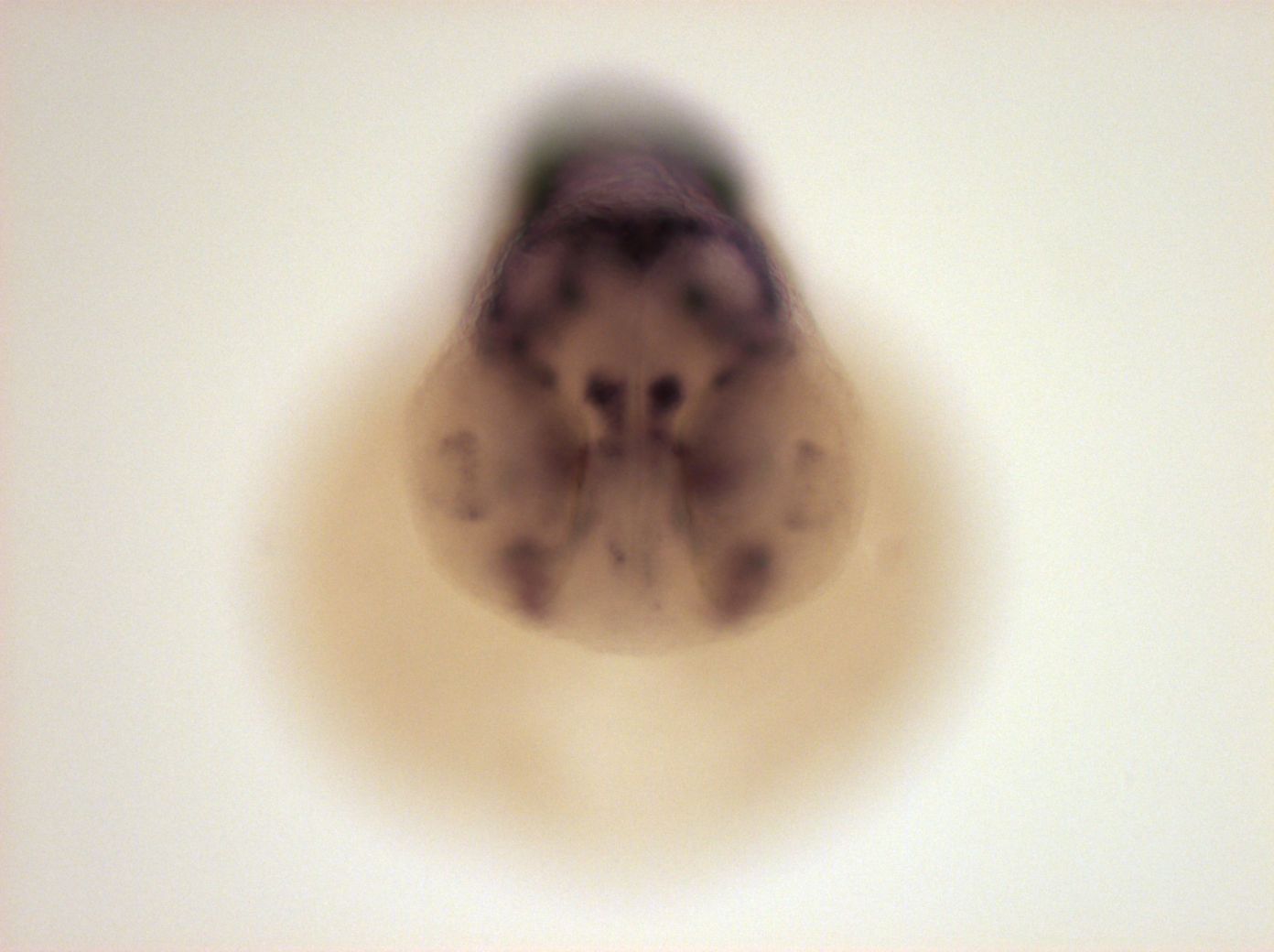
Figure Caption
Fig. 5 nose, epiphysis, ventral posterior tegmentum, dorsal cerebellum, dorsal hindbrain, cranial ganglia, nuclei in ventral hindbrain, ventral part of retina, habenula (left, very weak expression on right side), hypaxial part of anterior somites, spinal chord neurons.
Orientation
| Preparation | Image Form | View | Direction |
| whole-mount | still | frontal | anterior to top |
Figure Data