
Image
|
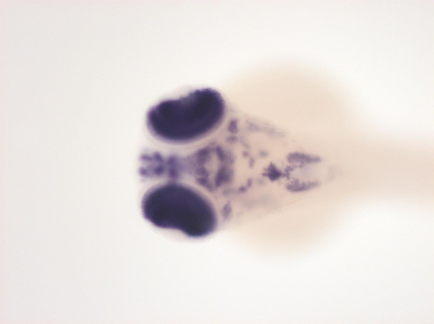
Figure Caption
Fig. 6 ventral telencephalon, hypothalamus, anterior ventral diencephalon, ventral anterior retina, trigeminal ganglion, pharyngeal arches, pancreas, ventral hindbrain and spinal chord neurons, dorsal spinal chord neurons, ganglion cell layer of retina (expression is stronger in ventral spinal chord)
Orientation
| Preparation | Image Form | View | Direction |
| whole-mount | still | dorsal | anterior to left |
Figure Data