
IMAGE
Fig. 6
- ID
- ZDB-IMAGE-020319-110
- Genes
- Publication
- Thisse et al., 2001 - Expression of the zebrafish genome during embryogenesis (NIH R01 RR15402)
- All Figures
- Figures for Thisse et al., 2001[cb110]
Image
|
Figure Caption
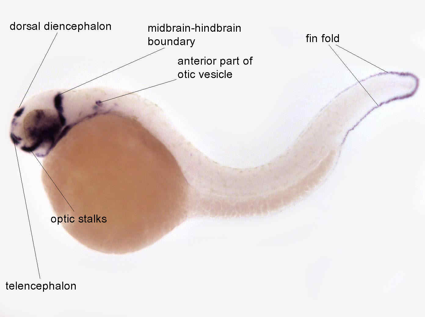
Fig. 6 At 36 hrs, expression is observed in the ganglion cell layer of the retina, the adenohypophysis, and the hyoid arch. Expression has disappeared from the posterior otic vesicle and the somites. An increase of expression is observed in median fin fold
Developmental Stage
Prim-15 to Prim-25
Orientation
| Preparation | Image Form | View | Direction |
| whole-mount | still | side view | anterior to left |
Figure Data
|
Anatomy Terms:
diencephalon
,
optic stalk
,
midbrain hindbrain boundary
,
otic vesicle
,
telencephalon
,
median fin fold
|