- Title
-
Unique developmental trajectories and genetic regulation of ventricular and outflow tract progenitors in the zebrafish second heart field
- Authors
- Paffett-Lugassy, N., Novikov, N., Jeffrey, S., Abrial, M., Guner-Ataman, B., Sakthivel, S., Burns, C.E., Burns, C.G.
- Source
- Full text @ Development
|
Identification of ZsYellow+ head muscles and endothelium in Tg(nkx2.5:ZsYellow) larvae. (A-F) Confocal z-stacks of pharyngeal regions in 72 hpf Tg(nkx2.5:ZsYellow) zebrafish larvae double immunostained to detect ZsYellow fluorescent protein and striated muscle (MF20 antibody) imaged in the green (pseudocolored yellow) and red channels, respectively. (G-I) Confocal z-stacks of the pharyngeal region in a 96 hpf Tg(nkx2.5:ZsYellow); Tg(fli1a:GFP) larvae double immunostained to detect ZsYellow and green fluorescent protein (GFP) imaged in the red (pseudocolored yellow) and green (pseudocolored cyan) channels, respectively. Single-channel (A,B,D,E,G,H) and merged (C,F,I) images are shown. Ventral (A-C,G-I) and lateral (D-F) views are shown. Anterior is upwards (A-C,G-I) or leftwards (D-F). For both experiments, little to no variability in the staining patterns was observed in the greater than 30 animals examined. Ventral pharyngeal arch (PA) 1 (mandibular) muscle: ima, intermandibular anterior. Middle PA1 muscles: imp, intermandibular posterior; am, adductor mandibulae. Dorsal PA1 muscles: lap, levator arcus palatine; do, dilator operculi. Ventral PA2 (hyoid) muscle: ih, interhyal. Middle PA2 muscle: hh, hyohyal. Dorsal PA2 muscles: ah, adductor hyomandibulae; ao, adductor operculi; lo, levator operculi. Vessel: ha, hypobranchial artery. Cardiac structures: oft, outflow tract; pp, parietal pericardium; v, ventricle. Scale bars: 25 µm. |
|
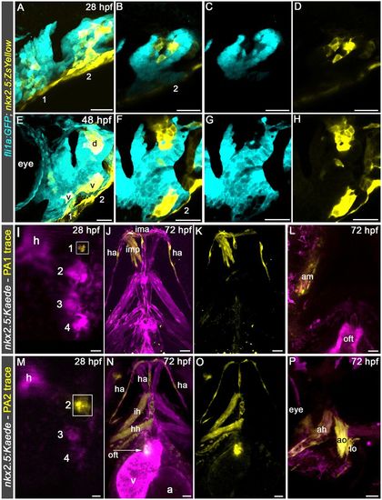
Visualization and prospective lineage tracing of head muscle and hypobranchial artery endothelial precursors in pharyngeal arches 1 and 2. (A-H) Confocal images of pharyngeal arch (PA) 1 and PA2 in 28 hpf (A-D) and 48 hpf (E-H) Tg(nkx2.5:ZsYellow); Tg(fli1a:GFP) embryos double immunostained for GFP and ZsYellow imaged in the green (pseudocolored cyan) and red (pseudocolored yellow) channels, respectively. Single planes (B-D,F-H) from merged confocal z-stacks (A,E) are shown as merged (B,F) or single-channel (C,D,G,H) images. Left lateral views are shown. Anterior is towards the left. Little to no variability was observed in greater than 30 animals examined. (I,M) Merged confocal z-stacks of pharyngeal regions in live 28 hpf Tg(nkx2.5:Kaede) embryos immediately following unilateral photoconversion of Kaede+ cells (boxed regions) in PA1 (I) or PA2 (M). (J,K,N,O) Confocal z-stacks of pharyngeal regions in the same embryos at 72 hpf shown as merged (J,L,N,P) or single-channel (K,O) images. All five animals with PA1 photoconversion demonstrated tracing to the ipsilateral PA1 head muscles and bilateral HA endothelium. Labeling of the intermandibular anterior muscle crossed the midline in some cases. All 14 animals with PA2 photoconversion demonstrated tracing to the ipsilateral PA2 head muscles, ipsilateral OFT and bilateral HA endothelium. Animals were imaged in the green (pseudocolored magenta) and red (pseudocolored yellow) channels. Dorsal (I,M), ventral (J,K,N,O) and lateral (L,P) views are shown. Anterior is upwards (I-K,M-O) or leftwards (L,P). Numbers label the pharyngeal arches. d, dorsal cluster; v, ventral cluster. Ventral pharyngeal arch (PA) 1 (mandibular) muscle: ima, intermandibular anterior. Middle PA1 muscles: imp, intermandibular posterior; am, adductor mandibulae. Ventral PA2 (hyoid) muscle: ih, interhyal. Middle PA2 muscle: hh, hyohyal. Dorsal PA2 muscles: ah, adductor hyomandibulae; ao, adductor operculi; lo, levator operculi. Vessel: ha, hypobranchial artery. Cardiac structures: oft, outflow tract; a, atrium; v, ventricle. Scale bars: 25 µm. |
|
Pharyngeal arch 2 muscles and hypobranchial artery endothelium derive from nkx2.5+ progenitors in the ALPM. (A-F) Confocal z-stacks (A,B,D,E) and schematic diagrams (C,F) of a 14-somite stage (ss) (16 hpf) Tg(nkx2.5:Kaede) embryo immediately following left-side photoconversion of the Kaede+ ALPM (anterior half; A-C) and the same embryo at 28 hpf (D-F) imaged in the green (pseudocolored magenta) and red (pseudocolored yellow) channels. Dorsal views are shown. Anterior is upwards. Merged (A,D) and single-channel (B,E) images are shown. In all five embryos, photoconverted cells traced to the heart and the pharyngeal arches as shown. (G-L) Confocal z-stacks of pharyngeal regions in 72 hpf Tg(nkx2.5:CreERT2), Tg(ubi:Switch) embryos pulsed with 4-OHT between tailbud and 16 ss (10-17 hpf). Animals were double immunostained to detect the mCherry reporter protein and striated muscle (MF20 antibody) before imaging in the red (pseudocolored yellow) and far-red (pseudocolored red) channels, respectively. Ventral (G-I,L) and left lateral (J,K) views are shown. Anterior is upwards (G-I,L) or leftwards (J,K). All 12 animals exhibited lineage tracing to the described structures. Numbers label the pharyngeal arches. h, heart. Ventral pharyngeal arch (PA) 1 (mandibular) muscle: ima, intermandibular anterior. Middle PA1 muscles: imp, intermandibular posterior; am, adductor mandibulae. Dorsal PA1 muscles: lap, levator arcus palatine; do, dilator operculi. Ventral PA2 (hyoid) muscle: ih, interhyal. Middle PA2 muscle: hh, hyohyal. Dorsal PA2 muscles: ah, adductor hyomandibulae; ao, adductor operculi; lo, levator operculi. Vessel: ha, hypobranchial artery. Cardiac structures: oft, outflow tract; v, ventricle. Scale bars: 25 µm. |
|
Unique trajectories of SHF ventricular and OFT progenitors. (A) Confocal z-stack of the pharyngeal region in a 28 hpf Tg(nkx2.5:Kaede) embryo immediately following bilateral photoconversion of Kaede+ cells in pharyngeal arch (PA) 2. A dorsal view is shown. Anterior is upwards. (B) Confocal z-stack of the ventricle and OFT in the same embryo at 72 hpf. The arrow highlights OFT cells carrying the photoconverted Kaede protein. All nine animals contained photoconverted cells in the OFT. (C,D) High-magnification confocal z-stacks of the OFT in a 72 hpf Tg(nkx2.5:Kaede) animal in which Kaede+ cells in PA2 were photoconverted bilaterally at 28 hpf. (E,F) Confocal z-stacks of a 21 hpf Tg(nkx2.5:Kaede) embryo immediately following photoconversion of Kaede+ cells in the cardiac cone. A dorsal view is shown. Anterior is upwards. (G,H) Confocal z-stack of the ventricle and OFT in the same embryo at 72 hpf. All 12 animals contained photoconverted cells in the ventricle but not in the OFT. All animals in A-H were imaged in the green (pseudocolored magenta) and red (pseudocolored yellow) channels. Merged (A-C,E,G) and single-channel (D,F,H) images are shown. Cardiac structures: h, heart; oft, outflow tract; v, ventricle. Scale bars: 25 µm. |
|
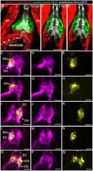
PA2 progenitors give rise to three OFT lineages. (A,B) Confocal images of the OFT in a 72 hpf Tg(fli1a:nGFP) embryo triple immunostained to detect OFT myocardium (MF20 antibody, red), smooth muscle (α-Eln2 antibody, green) and endothelium (α-GFP antibody, white). A z-stack (A) and single planes (B,B′) are shown. B′ is a duplicate of B without the labels and provides an unimpeded view of the physical relationships between OFT lineages. OFT endocardium is surrounded by two collars of muscle, a myocardial collar abutting the ventricle and an adjacent smooth muscle collar that extends to the ventral aorta. (C-Q) Single planes from confocal z-stacks of OFTs in 72 hpf live Tg(nkx2.5:Kaede) embryos in which right-sided (C-H,L-N), left-sided (I-K) or bilateral (O-Q) photoconversion of Kaede+ cells in PA2 was performed at 28 hpf. OFT cells carrying the native (pseudocolored magenta) and photoconverted (pseudocolored yellow) Kaede protein are shown. Merged (C,F,I,L,O) and single-channel (D,E,G,H,J,K,M,N,P,Q) images are shown. The lineage identities of the labeled cells (arrows) were inferred based on location. Ventral views, anterior up. sh, sternohyoideus muscle; va, ventral aorta; oft, outflow tract; SM, smooth muscle; EC, endocardium; CM, cardiomyocytes. Scale bars: 25 µm. |
|
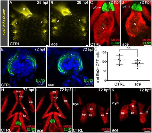
SHF ventricular and OFT progenitors are uniquely sensitive to perturbations in Fgf8a signaling. (A,B) Compound microscopic images of live 28 hpf control (CTRL) sibling (A) or ace (B, n=33) embryos carrying the nkx2.5:ZsYellow transgene. Dorsal views are shown. Anterior is upwards. The dotted lines demarcate the arterial pole of the heart tube. The asterisk highlights extra-cardiac ZsYellow+ anterior SHF progenitors absent in ace mutants. All 74 control and all ace animals displayed the ZsYellow distributions shown. (C,D) Confocal z-stacks of the ventricle and OFT in 72 hpf CTRL and ace animals double immunostained to detect striated muscle (MF20 antibody) and Eln2+ smooth muscle OFT cells imaged in the red and green channels, respectively. All 76 control and all 25 ace animals contained Eln2+ OFTs. Ventral views. Anterior is upwards. (E,F) Confocal z-stacks of the OFTs in 72 hpf CTRL and ace mutant embryos immunostained to visualize Eln2 (green) and counterstained with DAPI (blue) to detect nuclei. White dots highlight nuclei in cells surrounded by Eln2. (G) Dot plot showing the quantification of Eln2+ OFT cells in 72 hpf control and ace mutant embryos. An unpaired t-test was used to evaluate statistical significance. ns, not significant. (H-K) Confocal z-stacks of pharyngeal regions in 72 hpf control and ace animals double immunostained to detect striated muscle (MF20 antibody) and Eln2+ smooth muscle OFT cells imaged in the red and green channels, respectively. All 76 CTRL and 14/25 ace animals contained the head muscles in the pattern shown. Ventral (H,I) and lateral (J,K) views are shown. Anterior is upwards (H,I) or leftwards (J,K). Number two labels pharyngeal arch 2. h, heart. Ventral pharyngeal arch (PA) 1 (mandibular) muscle: ima, intermandibular anterior. Middle PA1 muscles: imp, intermandibular posterior; am, adductor mandibulae. Dorsal PA1 muscles: lap, levator arcus palatine; do, dilator operculi. Ventral PA2 (hyoid) muscle: ih, interhyal. Middle PA2 muscle: hh, hyohyal. Dorsal PA2 muscles: ah, adductor hyomandibulae; ao, adductor operculi; lo, levator operculi. Cardiac structures: oft, outflow tract; v, ventricle. Scale bars: 25 µm. |
|
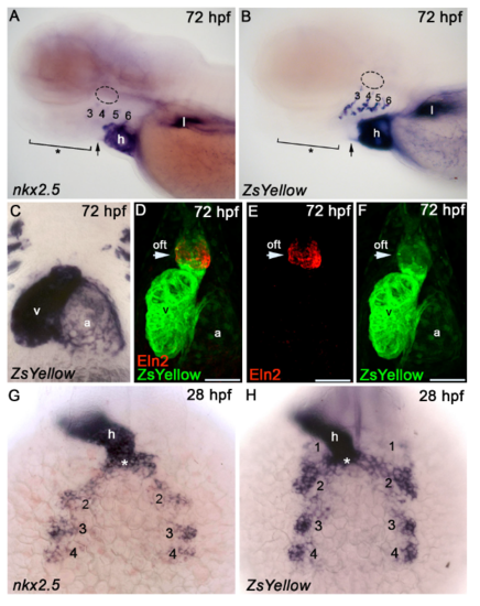
Expression analysis of nkx2.5 and Zs yellow in wild type and Tg(nkx2.5:Zs Yellow) animals. (A,B) Lateral views of 72 hours post fertilization (hpf) wild type (A) or Tg(nkx2.5:Zs Yellow) (B) zebrafish embryos processed for in situ hybridization with nkx2.5 or ZsYellow riboprobes, respectively. Anterior to let. The brackets(*) highlight the locations of ventral and middle pharyngeal arch (PA) 1 and PA2 muscles as well as hypobranchial artery endothelium. The circles drawn with dotted lines show the location of dorsal PA1 and PA2 muscles. The arrows highlight the location fo the OFT, which is negative for nkx2.5(A) or ZsYellow (B) transcripts. The signals located dorsal to the heart in (B) are from cells associated with th pharyngeal arch arteries. (C-F) Ventral views of the heart and OFT in 72 hpf Tg(nkx2.5:ZsYellow) animals processed for in situ hybridization with a ZsYellow riboprobe (C) or double immunostained to detect ZsYellow (green) and Eln2+OFT smooth muscle cells (red; D-F). Anterior up. Merged (D) and single channel (E,F) confocal Z-stack are shown. The arrows highlight the ZsYellow+, Eln2+ OFT that protrudes from the ventricle, which is negative for ZsYellow transcripts at this stage(C). (G,H) Dorsal views of 28 hpf wild type (G) or Tg(nkx2.5:ZsYellow) (H) zebrafish embryos processed for in situ hybridization with nkx2.5 or ZsYellow riboprobes, respectively. The asterisk (*) highlights SHF progenitors for the distal ventricular myocardium. Little to no variability was observed in the greater than 30 animals examined. Numbers label the pharyngeal arches. Abbreviations: h, heart; l, liver; v, ventricle; a, atrium; oft, outflow tract. Scale Bars=50µm. |
|
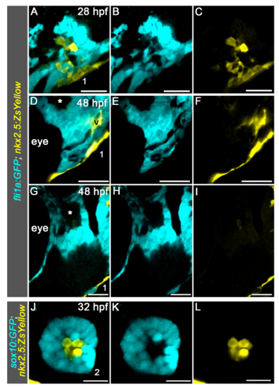
Visualization of head muscle and hypobranchial endothelial progenitors in the mandibular arch. (A-I) Singles planes from confocal Z-stacks shown in Figure 2A (A-C) and Figure 2E (D-I). The (*)s in (D,G) highlight the dorsal mesodermal core of pharyngeal arch (PA) 1, which is devoid of ZsYellow+ cells. Lateral views, anterior left. (J-L) Single plane from confocal Z-stack of PA2 in 32 hours post fertilization (hpf) Tg(sox10:gfp); Tg(nkx2.5:ZsYellow) double transgenic embryo. Dorsal views, anterior up. For both experiments, 3/3 animals exhibited ZsYellow fluorescence in the described locations. Numbers label the pharyngeal arches; Abbreviations: v, ventral cluster. Scale bars=25µm. |
|
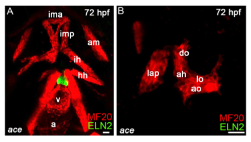
Analysis of head muscles in ace embryos. (A,B) Confocal Z-stacks of ace mutant embryos double immunostained to detect skeletal muscle (MF20 antibody) and Eln2+OFT cells imagined in the red and green channels, respectively. Representative example of ace mutant embryos containing mispatterned head muscles in pharyngeal arch 1 and 2(n=11/25). Ventral views, anterior up (A). Lateral view, anterior left (B). Please see Figure 1 legend for HM abbreviations. Scale bars=25µm |


