
Fig. S2
- ID
- ZDB-FIG-180501-28
- Publication
- Paffett-Lugassy et al., 2017 - Unique developmental trajectories and genetic regulation of ventricular and outflow tract progenitors in the zebrafish second heart field
- Other Figures
- All Figure Page
- Back to All Figure Page
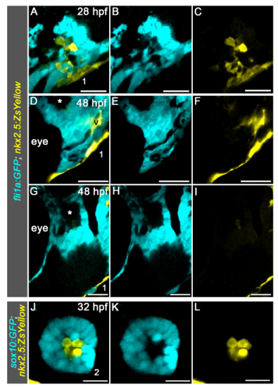
Visualization of head muscle and hypobranchial endothelial progenitors in the mandibular arch. (A-I) Singles planes from confocal Z-stacks shown in Figure 2A (A-C) and Figure 2E (D-I). The (*)s in (D,G) highlight the dorsal mesodermal core of pharyngeal arch (PA) 1, which is devoid of ZsYellow+ cells. Lateral views, anterior left. (J-L) Single plane from confocal Z-stack of PA2 in 32 hours post fertilization (hpf) Tg(sox10:gfp); Tg(nkx2.5:ZsYellow) double transgenic embryo. Dorsal views, anterior up. For both experiments, 3/3 animals exhibited ZsYellow fluorescence in the described locations. Numbers label the pharyngeal arches; Abbreviations: v, ventral cluster. Scale bars=25µm. |