
FIGURE 4
- ID
- ZDB-IMAGE-260703-56
- Antibodies
- Publication
- Wang et al., 2026 - Atypical Cadherin Fat2 is Involved in Axogenesis of Cerebellar Granule Cells in Zebrafish
- All Figures
- Figures for Wang et al., 2026
|
FIGURE 4
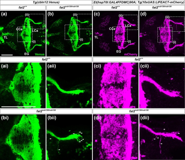
Granule cell morphology and Actin organization in wild‐type and fat2 mutant larvae. (a–d) Expression of Venus in 5‐dpf Tg(cbln12:Venus) larvae in wild‐type (a) and fat2nub128/nub128 backgrounds (b) (n = 5 for each background), and expression of mCherry in 5‐dpf Et(hsp70l:GAL4FFDMC)nkhspGFFDMC90AEt; Tg(10xUAS:LIFEACT‐mCherry) larvae in wild‐type (c) and fat2nub128/nub128 backgrounds (d) (n = 5 for each background). Signals were detected by immunostaining. Dorsal views with anterior to the left. (ai–di) Higher magnification views of the corpus cerebelli. (aii–dii) Higher magnification views of granule cell axons in the caudolateral lobes. Arrowheads indicate branched bundles. Scale bar: 100 μm in (a) (applies to a–d) and (aa) (applies to ai–di and aii–dii). Regions of the cerebellum, including CCe, EG, LCa, and TL, are indicated in the images. Images are representative of the samples examined, and the observed phenotypes were consistent across all samples in each condition (n = 5).