
FIGURE 3
- ID
- ZDB-IMAGE-260626-16
- Publication
- Rezi et al., 2026 - ZDHHC5 interacts physically and functionally with DLG1 at primary cilia and regulates ciliary length and kidney morphology
- All Figures
- Figures for Rezi et al., 2026
|
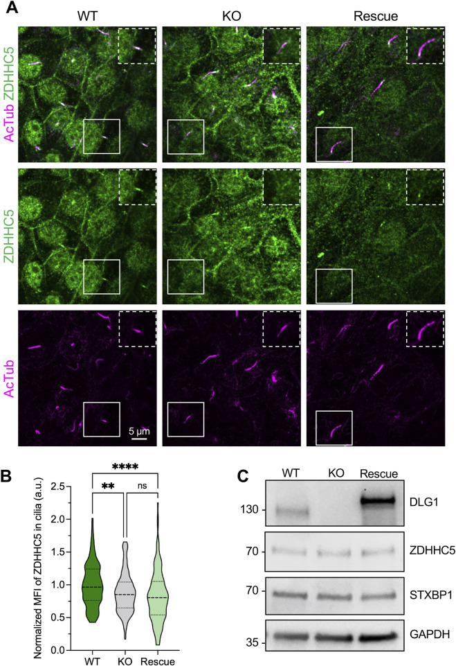
FIGURE 3
Ciliary localization of ZDHHC5. (A) IFM analysis of ciliary localization of ZDHHC5 in control (WT) and Dlg1 knockout cells without (KO) or with (Rescue) stable re-expression of mCherry-DLG1. Cells were cultured for 24 h in supplement-free medium and subjected to IFM analysis using antibodies against ZDHHC5 (green) and acetylated α-tubulin (AcTub; magenta) to stain the ciliary axoneme. Insets show zoom-ins of the cilium-basal body axis. Note that average ciliary length is unaffected by DLG1 loss under these conditions (