IMAGE

Fig. 2

ID
ZDB-IMAGE-260515-35
Publication
Lim et al., 2026 - Functional impact of the ATP1A3-p.A813V variant: insights into a calcium-driven hyperexcitability cascade in rapid-onset dystonia-Parkinsonism
All Figures
Figures for Lim et al., 2026
Image
Figure Caption

Fig. 2

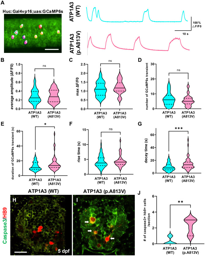
ATP1A3-p.A813V mutation induces prolonged Ca2+ transients, leading to neuronal apoptosis. (a) Representative image of the spinal cord in Tg(Huc:Gal4;uas:GCaMP6s);nacre zebrafish at 3 days post-fertilization (DPF). Regions of interest (ROIs) designated 10 neurons per zebrafish. Scale bar: 50 μm. Individual traces depict GCaMP6s fluorescence signals from spinal neurons overexpressing ATP1A3 (blue) and those overexpressing ATP1A3-p.A813V (pink). (b–g) Quantification of various parameters of GCaMPs transients in neurons. (b) The average amplitude of GCaMPs transients measured in neurons. Mann–Whitney test; n = 18 and n = 16, respectively. (c) Quantification of the maximum amplitude of GCaMPs transients. Unpaired t-test; n = 18 and n = 16, respectively. (d) Quantification of the number of GCaMPs transients. Mann–Whitney test; n = 19, n = 18, and n = 16, respectively. (e) Quantification of duration of GCaMPs transients. Unpaired t-test; n = 18 and n = 16, respectively. (f) Quantification of the rise time of GCaMPs transients. Mann–Whitney test; n = 18 and n = 16, respectively. (g) Quantification of the decay time of GCaMPs transients. Mann–Whitney test; n = 18 and n = 16, respectively. (h, i) representative transverse section images of the spinal cord in zebrafish overexpressing ATP1A3 (WT) (h) and ATP1A3 (p.A813V) (i) at 5 DPF. Immunohistochemistry labeling with anti-active caspase3 antibody (green) in Tg(Hb9:mCherry) zebrafish revealing neuronal cell death. Scale bar: 10 μm. (j) Quantification of Caspase3+ Hb9+ neurons per section at 5 DPF. Mann–Whitney test. analysis conducted on n = 8 zebrafish per group. Statistical significance denoted as not significant (n.s.), *p < 0.05, **p < 0.01, ***p < 0.001

Acknowledgments
This image is the copyrighted work of the attributed author or publisher, and ZFIN has permission only to display this image to its users. Additional permissions should be obtained from the applicable author or publisher of the image. Full text @ J Transl Med