
FIGURE 2
- ID
- ZDB-IMAGE-260327-13
- Genes
- Publication
- Chen et al., 2026 - Sirt1 coordinates the mitochondrial UPR and myocellular proteostasis to preserve muscle integrity during muscle atrophy in zebrafish
- All Figures
- Figures for Chen et al., 2026
|
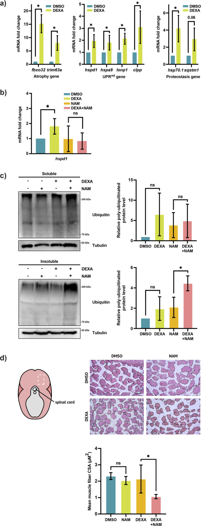
FIGURE 2 Sirt1 and the UPRmt promote myocellular proteostasis and muscle maintenance. (a) Left: qRT-PCR analysis of the skeletal muscle from DEXA-treated adult zebrafish showing the expression of the atrophy genes fbxo32 and trim63a (*p < 0.05, t-test, two-tailed, error bar indicates s.d.). Middle: qRT-PCR analysis of the skeletal muscle from DEXA-treated adult zebrafish showing the expression of the UPRmt genes hspd1, hspa9, lonp1, and clpp (*p < 0.05, t-test, two-tailed, error bar indicates s.d.). Right: qRT-PCR analysis of the skeletal muscle from DEXA-treated adult zebrafish showing the expression of the proteostasis genes hsp70.1 and sqstm1 (*p < 0.05, t-test, two-tailed, error bar indicates s.d.). (b) qRT-PCR analysis of the skeletal muscle from adult zebrafish treated with DEXA and/or nicotinamide (NAM) showing the expression of the UPRmt gene hspd1 (*p < 0.05, t-test, two-tailed, error bar indicates s.d.). (c) Western blot and quantification of poly-ubiquitinated proteins in the detergent-soluble and -insoluble fractions of the protein lysates from the skeletal muscle of adult zebrafish treated with DEXA and/or NAM (*p < 0.05, t-test, two-tailed, error bar indicates s.d.). (d) Representative light microscopy images of hematoxylin and eosin (H&E)-stained cross-sections of the skeletal muscle from adult zebrafish treated with DEXA and/or NAM. The images are acquired from the general area of the adult fish musculature indicated by the white rectangle in the diagram to the left. Scale bar represents 100 µm. Histogram showing the average cross-sectional area of the skeletal muscle fibers from adult zebrafish treated with DEXA and/or NAM (*p < 0.05, t-test, two-tailed, error bar indicates s.d.).