
Image
|
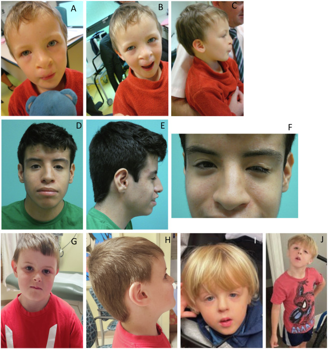
Figure Caption
Fig. 2 Photographs of probands with de novo variants in EIF3B or EIF3A (A–C) Clinical images of proband #4, with a canonical acceptor splice site variant in EIF3B; (D–F) proband #9 with a loss-of-function variant in EIF3B; (G–H) proband #15, with a loss-of-function variant EIF3A; and (I and J) proband #18, with a loss-of-function variant in EIF3A.
Acknowledgments
This image is the copyrighted work of the attributed author or publisher, and
ZFIN has permission only to display this image to its users.
Additional permissions should be obtained from the applicable author or publisher of the image.
Full text @ Am. J. Hum. Genet.