
|
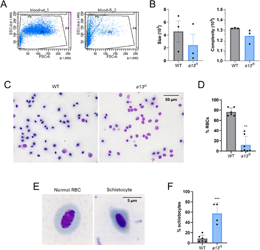
Fig. 1 Depletion of erythrocytes and presence of schistocytes in the peripheral blood of adamts13i5 mutants. (A) flow cytometry analysis of the peripheral blood of adult zebrafish. (B) Analysis of the size (left) and complexity (right) of WT (grey) and a13i5 (blue) fish. (C) Representative peripheral blood smears of the erythrocytes. (D) Quantification of the RBCs in the peripheral blood of adult zebrafish: WT (grey), a13i5 (blue). (E) Illustrative comparison between a normal RBC and a schistocyte. (F) Quantification of SCs in the peripheral blood of adult zebrafish: WT (grey), a13i5 (blue). Data are shown as means±s.d. of 3-7 biological replicates. Each dot represents a single adult fish. **P≤0.01 and ***P≤0.001 by two-tailed Student's t-test. Blood smear images were obtained using a 63× oil-immersion objective.