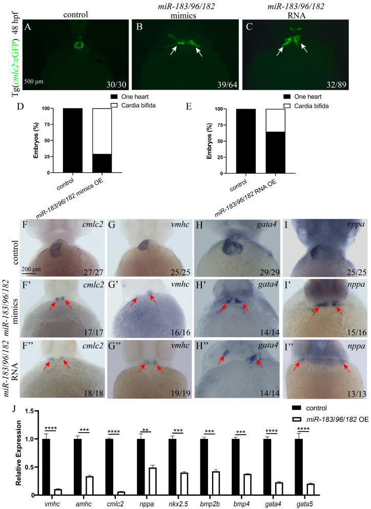
Co-expression of the miR-183 family leads to abnormal migration of myocardial precursor cells. (A–C) Fluorescence images of the hearts of Tg (cmlc2: eGFP) transgenic zebrafish embryos at 48 hpf (ventral view). Control embryos exhibit a normally fused heart tube (A) (green). Both miR-183/96/182 mimics OE (B) and miR-183/96/182 capped RNA OE (C) embryos exhibit cardia bifida (white arrow heads indicate separate cardiac primordia). (D,E) Quantification of cardia bifida in miR-183 cluster mimics OE (D) and miR-183/96/182 capped RNA OE (E) embryos. (F–I) WISH detected cmlc2 (F,F’,F”), vmhc (G,G’,G”), gata4 (H,H’,H”), and nppa (I,I’,I”) in embryos of the 48 hpf control, miR-183/96/182 mimics OE and miR-183/96/182 capped RNA OE (red arrow heads indicate separate cardiac primordia). (J) The mRNA expression levels of heart development-related genes in control, miR-183/96/182 mimics OE embryos were analyzed by RT-qPCR at 42 hpf. The number at the bottom right indicates the number of embryos. ** p < 0.01, *** p < 0.001, **** p <0.0001.
Acknowledgments
This image is the copyrighted work of the attributed author or publisher, and
ZFIN has permission only to display this image to its users.
Additional permissions should be obtained from the applicable author or publisher of the image.
Full text @ Biomolecules
Your Input Welcome
Thank you for submitting comments. Your input has been emailed to ZFIN curators who may contact you if
additional information is required.
Oops. Something went wrong. Please try again later.