
|
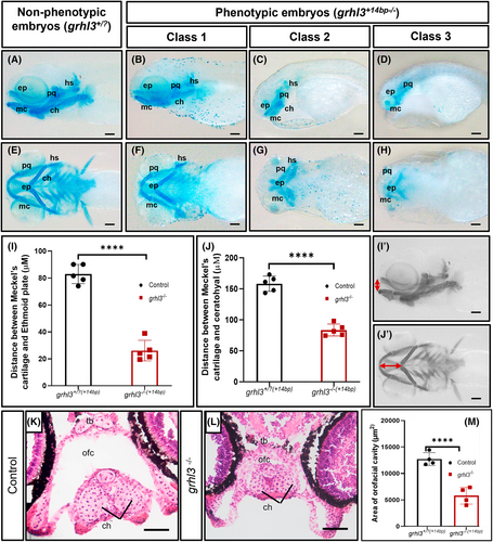
Fig. 4 Craniofacial development in grhl3−/−(+14 bp) embryos at 4 dpf. (A–H) Class 1, class 2, and class 3 grhl3−/−(+14bp) embryos and non-phenotypic siblings (4 dpf) stained with Alcian blue, showing lateral (A–D) and ventral (E–H) views of the craniofacial cartilage (palatoquadrate [pq], ceratohyal [ch] Meckel's cartilage [mc], ethmoid plate [ep], and hyosymplectic [hs]). (I-I') The height between Meckel's cartilage and ethmoid plate (indicated by vertical double-headed red arrow) and (J-J') the distance between Meckel's cartilage and ceratohyal (indicated by horizontal double-headed red arrow) were measured and analyzed in grhl3−/−(+14bp) embryos and non-phenotypic siblings. (K, L) H&E staining of transverse sections across the rostral region of 7 dpf class 1 grhl3−/− mutant and control embryos (ceratohyal [ch] trabecular bar, orofacial cavity [ofc]). (M) Orofacial cavity area of grhl3−/− and control embryos. Data are shown as mean ± SEM; n = 5. Statistical significance determined by Student's t-test ****p <.001. Scale bars: 100 μm (A-J'), 50 μm (K, L).