
|
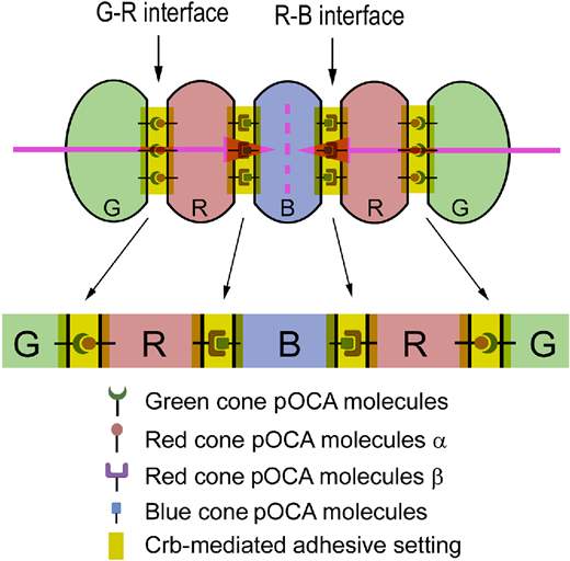
Fig. 7 The Lego self-assembly model for RGB cone pentamer formation and maintenance. Diagrams illustrate the hypothesized planar OCAs (pOCAs) that mediate G–R and R–B coalescence. Each type of RGB cone expresses a unique set of pOCA molecules. Blue-cone-specific pOCA molecules are distributed to two opposing parallel surfaces, where they bind to red-cone-specific pOCA molecules β to recruit two red cones in parallel flanking positions. Red cones also express another set of red-cone-specific pOCA molecules α, which localize opposite the pOCA molecules β and bind to green-cone-specific pOCA molecules, thus recruiting a green cone on the side opposite from the central blue cone. These parallel, anisotropically distributed, and cell-type-specific OCAs ultimately dictate the pentameric configuration. Crb2b localizes to all of these intercellular interfaces to play two roles: First, it assists in general adhesion by augmenting the adhesion between pentamer members; second, it helps to restrict G–R cone-specific pOCAs to the parallel junctional interfaces to prevent them from expanding to other membrane regions, which would result in square aggregation of green and red cones of neighboring pentamers. RGB cones are color coded; various OCA molecules are symbolized with distinct icons. The pink arrows indicate the opposing planar polarity within a pentamer, and the pink dashed line indicates the axis of the mirror symmetry of the planar polarities within a pentamer.