
|
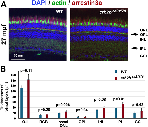
Fig. 6 The apicobasal polarization of the retina and photoreceptors remains largely intact in crb2bsa21178 homozygous mutants at 27 mpf. (A) Staining of actin (green, by phalloidin), nuclei (blue, by DAPI), and arrestin-3a (red, by the Zpr1 antibody) retinal layers: ONL, outer nuclear layer; OPL, outer plexiform layer; INL, inner nuclear layer; IPL, inner plexiform layer; GCL, ganglion cell layer. Images were taken from the central retinal regions. (B) The thicknesses of retinal layers of the wild-type and crb2bsa21178 homozygous mutants. O-I, distance between the outer limiting membrane and the inner limiting membrane; RGB, thickness of the RGB nuclear layer; basal ONL, thickness of the outer nuclear layer region that is basal to the outer limiting membrane. Student's two-tailed and paired t-tests were used to compare the differences in retinal layer thicknesses between the wild-type and mutant samples (n = 4 fish).