
|
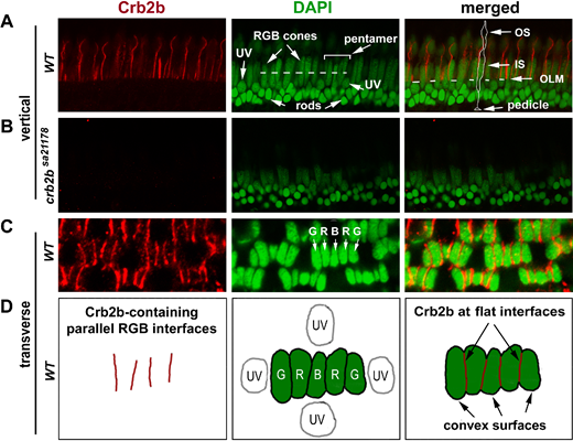
Fig. 2 Crb2b localization in the pentamers of RGB cones in 12-mpf retina. (A) Crb2b immunostaining (red) of radial sections of the wild-type photoreceptor layer. The DAPI staining (green) revealed the differences in staining intensities and positioning among the photoreceptor nuclei: lightly stained and more apical positioned long RGB cone nuclei; lightly stained round UV cone nuclei immediately basal to the outer limiting membrane (OLM, the dashed line in the merged panel); and intensively stained smallest rod nuclei at the basal half of the outer nuclear layer. The dashed line in the green panel indicates the position where pentamers were examined at the transverse plane (see panels C, D). A pentamer, flanked by two UV cones, is indicated by a bracket. In the merged image panel, an RGB cone is contoured to show its general polarized morphologies, with its pedicle, inner segment (IS), and outer segment (OS) indicated by arrows. (B) No Crb2b signals (red) were detected in homozygous crb2bsa21178 mutant retinas (radial section). (C, D) Crb2b immunostaining (red) of a transverse section of the photoreceptor layer revealed that Crb2b localized to the junctional interfaces between RGB cones of pentamers, as depicted by the diagrams in D. Note the lack of Crb2b signals at the adjacent convex cell surfaces.