
Fig. 2
- ID
- ZDB-IMAGE-251016-27
- Publication
- Yousaf et al., 2025 - Bi-allelic deleterious variants in SNAPIN, which encodes a retrograde dynein adaptor, cause a prenatal-onset neurodevelopmental disorder
- All Figures
- Figures for Yousaf et al., 2025
|
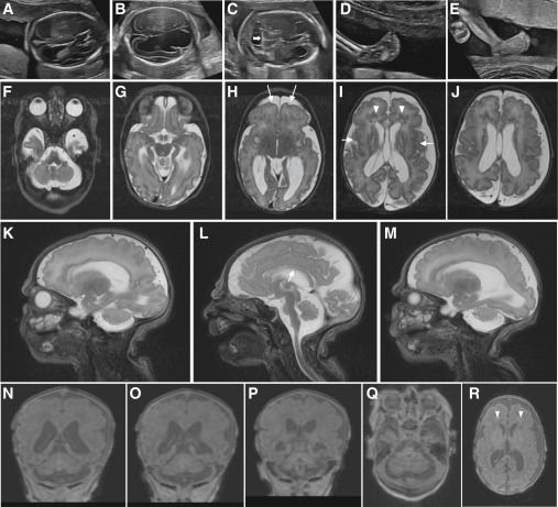
Fig. 2 Clinical imaging of the family B and family E probands who harbor bi-allelic variants in SNAPIN (A–E) Prenatal ultrasound imaging of the family B-II:3 fetus showing progression of ventriculomegaly at (A) 19 to (B) 21 weeks of gestation, with disruption of ventral parenchyma at 21 weeks; (C) cerebellar hypoplasia at 21 weeks of gestation (white horizontal arrow); and (D and E) progression of clubfeet at (D) 19 to (E) 21 weeks. (F–R) Brain MRI of the family E-II:3 proband, born at 38 weeks of gestation, at 3 days of age showing a complex brain malformation including microcephaly with simplified gyral pattern (arrows, H), polymicrogyria (arrows, I) (G–R), thin corpus callosum (arrow, L), bilateral enlargement of lateral ventricles (H–J, N, and O), prominent periventricular white matter lesions suggestive of mixed subcortical heterotopias (arrow heads, I and R), and pronounced pontocerebellar hypoplasia (F, L, and Q). (F–J) T2-weighted axial images, (Q and R) T1-weighted axial images, (K–M) T2-weighted sagittal images, and (N–P) T1-weighted coronal images.