
Fig. 5
- ID
- ZDB-IMAGE-250917-50
- Publication
- Leong et al., 2025 - RAB23 loss-of-function mutation causes context-dependent ciliopathy in Carpenter syndrome
- All Figures
- Figures for Leong et al., 2025
|
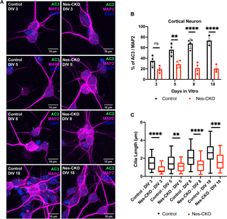
Fig. 5 Rab23 plays critical roles in neuronal primary cilia formation and elongation. (A) Representative co-immunocytochemistry images depict the process of primary cilia elongation in the control and Nes-CKO primary cortical neuron cultures at DIV 3 to DIV18. Neurons were co-immunostained for MAP2 (magenta), and the primary cilia marker AC3 (green) respectively. (B) Graph depicts the percentage of ciliation, i.e., quantification of the proportion of ciliated neurons (AC3 + MAP2+) in the MAP2 positive neurons. Data represents three to four independent experiments. n.s. = not significant, ** P value 0.01, **** P-value ≤ 0.0001, two-way ANOVA. (C) Graph depicts the measurement of cilia length of primary cortical neurons cultures at DIV 3 to DIV18. Box plot represents data of ~70–80 cilia in each group obtained from three to four independent experiments. ** P value 0.01, *** P value 0.001, **** P-value ≤ 0.0001, two-way ANOVA.