
Fig. 4
- ID
- ZDB-IMAGE-250917-49
- Publication
- Leong et al., 2025 - RAB23 loss-of-function mutation causes context-dependent ciliopathy in Carpenter syndrome
- All Figures
- Figures for Leong et al., 2025
|
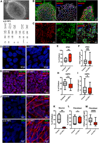
Fig. 4 Carpenter syndrome patient-derived iPSC-differentiated neurons exhibit severely impaired ciliogenesis. (A-B) Fibroblasts isolated from skin biopsy from a Carpenter syndrome patient bearing the RAB23 biallelic p.(L145*) variant was reprogrammed into iPSC clones. (A) Top: A representative phase contrast image showing successfully reprogrammed-iPSC clone displaying a normal karyotype (bottom), and pluripotency characterized by positive (B) co-immunostainings of human stem cell markers SSEA4, NANOG, OCT3/4 and TRA-1-60 respectively. (C) Representative co-immunocytochemistry images depicting positive expression of NESTIN (red) and SOX2 (green) on neural progenitor stem cells induced from iPSCs. (D) Representative co-immunocytochemistry images depicting the iPSCs of healthy adults (control) and Carpenter syndrome patients (p.(L145*)). Cells were immunostained for the primary cilia marker ARL13B (red). (E) Graph depicts the quantification of the percentage of ciliation in iPSCs. Box plot represents data from four independent experiments. n.s. = not significant. Unpaired Student’s t-test. (F) Box plots depict the measurements of cilia length on iPSCs. Data represents the measurements of ~80-90 cilia in each genotype obtained from four independent experiments. ** P value 0.01. Unpaired Student’s t-test. (G) Representative co-immunocytochemistry images depicting healthy adult and Carpenter syndrome patient iPSC-derived human neural progenitor cells (hNPCs) co-immunostained for SOX2 (magenta), NESTIN (red) and primary cilia marker ARL13B (green). (H) Graph depicts the quantification of the percentage of ciliation in hNPCs. Box plot represents data from at least five independent experiments. n.s. = not significant. Unpaired Student’s t-test. (I) Box plot depicts the measurements of cilia length on hNPCs. Data represents the measurements of ~65–80 cilia in each genotype obtained from at least three independent experiments. *P value 0.05. Unpaired Student’s t-test. (J-K) Representative co-immunocytochemistry images depict MAP2+ (red) neurons differentiated from iPSCs of healthy adults (control) and Carpenter syndrome patients (p.(L145*)). Primary cilia were labelled by AC3 (green). (K) Graph depicts the percentage of ciliation, i.e., quantification of the proportion of ciliated neurons (AC3 + MAP2+) in the MAP2 positive neuronal population. A significant reduction in ciliation was observed in neurons bearing p.(L145*) variant. Box plot represents data from four independent experiments. ** P value 0.01 Unpaired Student’s t-test. (L) Graph depicts the quantification of the percentage of ciliation in primary fibroblast cells. The primary cilia in human fibroblast were visualised by immunostaining of acetylated-alpha-tubulin and ARL13B. Primary fibroblasts cultured from a healthy donor and a Carpenter syndrome patient with p.(L145*) variant show comparable percentages of ciliation. Box plot represents data from five independent experiments. n.s. = not significant. Unpaired Student’s t-test. (M) Box plot depicts the measurements of cilia length on fibroblast. Data represents the measurements of ~65–80 cilia in each group obtained from five independent experiments. ****P value 0.0001. Unpaired Student’s t-test.