
Fig. 3
- ID
- ZDB-IMAGE-250917-48
- Genes
- Antibodies
- Publication
- Leong et al., 2025 - RAB23 loss-of-function mutation causes context-dependent ciliopathy in Carpenter syndrome
- All Figures
- Figures for Leong et al., 2025
|
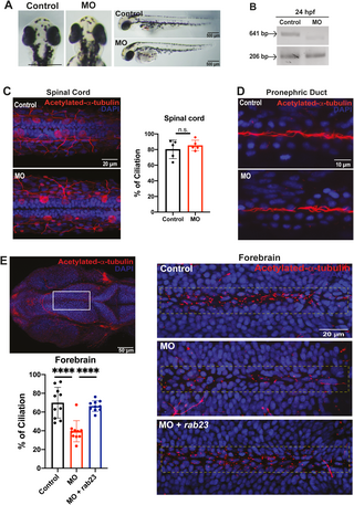
Fig. 3 Knockdown of rab23 in zebrafish affects primary cilia formation at the rostral brain ventricle. (A) Morpholino-mediated knockdown of rab23 in zebrafish. Bright-field dorsal view microscopy images of control and morphant (MO) at 72 hpf. (B) Representative gel image showing normal splicing of rab23 in control (641 bp) or inhibited splicing in morphants 24 hour-post-fertilization (hpf), resulting in less or shorter spliced product. Actin (206 bp) was used as the internal control. (C) Representative images and graph showing largely unaffected percentage of cilia lining the central canal of the spinal cord between control and morphant. n = 5 animals for each group. Error bars depict S.D. n.s. = not significant. Unpaired Student’s t-test. (D) Representative images showing largely unaffected cilia number at the central canal of the pronephric duct between control and morphant. (E) Representative images showing an obvious reduction of cilia number present in the forebrain ventricle of morphant as compared to control. Bottom right: rab23 morphant rescued by injecting mRNA of rab23 (bottom panel). Bottom left panel: Graph presenting quantitative analysis of cilia number in the brain ventricle of control, morphants and rescue group at 24 hpf, respectively. n = 10 for each group. Error bars depict S.D. **** P value 0.0001 One-way ANOVA.