
|
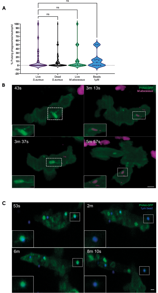
Fig. 3 Pulsatile bursts of PHAkt-eGFP recruitment are a neutrophil response to prey. (A) Violin plot showing the percentage of pulsing phagosomes per phagocytic neutrophil for live and dead S. aureus, live M. abscessus and 1 µm beads. Data shown are the median with the 25th and 75th percentiles. Live S. aureus, 46 phagosomes analysed from 11 independent larvae, from 11 experiments; dead S. aureus, 54 phagosomes, ten independent larvae, from ten experiments; live M. abscessus, 18 phagosomes, six independent larvae, six experiments; 1 µm beads, 11 phagosomes, four independent larvae, four experiments. ns, not significant (Kruskal–Wallis test). (B) ‘Pulses’ occur on phagosomes containing M. abscessus. PHAkt-eGFP recruits to phagosomes during phagocytosis (43 s) before gradually diminishing (3 min 13 s). PHAkt-eGFP then re-recruits to the phagosome (1st pulse; 3 min 37 s) and then diminishes again (5 min 57 s). Scale bar: 2 µm. (C) Pulses occur on phagosomes containing 1 µm beads. PHAkt-eGFP recruits to phagosomes during phagocytosis (53 s) before gradually diminishing (2 min). PHAkt-eGFP then re-recruits to the phagosome (1st pulse; 6 min) and then diminishes again (8 min 10 s). Phagocytosis starts at 0 min. Scale bar: 1 µm.