
|
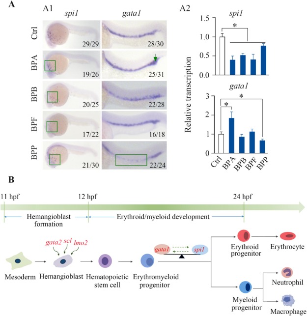
Fig. 5 Effects of BPA, BPB, BPF, and BPP on the differentiation of hemangioblasts during zebrafish development. WISH (A1) and qPCR (A2) analyses revealed the transcriptional levels of the spi1 and gata1 genes in embryos exposed to 1000 μg/L BPA, BPB, BPF, or BPP at 24 hpf. Schematic diagram of hematopoiesis during early zebrafish development showing that BPA, BPB, BPF, and BPP blocked the differentiation of hemangioblasts into myeloid progenitor cells through regulating spi1 and gata1 expression (B); significantly affected endpoints are marked in red. The number shown in the lower right-hand corner of each picture is the number of embryos exhibiting the typical phenotype shown in the picture to the total number of embryos observed. The qPCR results are shown as the means ± SD (n = 3). Significant differences between bisphenol-treated and control groups are denoted by asterisks (p < 0.05).