
Fig. 1
- ID
- ZDB-IMAGE-250909-70
- Genes
- Publication
- Robichon et al., 2025 - kcnb1 loss of function in zebrafish causes neurodevelopmental and epileptic disorders associated with γ-aminobutyric acid dysregulation
- All Figures
- Figures for Robichon et al., 2025
|
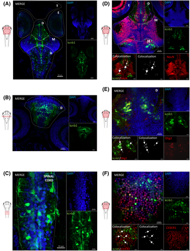
Fig. 1 kcnb1 is expressed in distinct cell subtypes and various regions of the central nervous system in 6 days post fertilization (dpf) wild-type (WT) zebrafish. (A, B) Horizontal and transversal sections of WT zebrafish expressing cells (4,6-diamidino-2-phenylindole [DAPI]; blue) labeled with anti-kcnb1 (green), showing a large expression of the protein in the central nervous system (CNS) at 6 dpf. The protein is expressed in the telencephalon, diencephalon, midbrain including the optic tectum, and hindbrain comprising the cerebellum and the spinal cord (A: scale bar = 50 μm, magnification = 10×; B: scale bar = 30 μm, magnification 20×). (C) Horizontal section of a WT zebrafish at 6 dpf showing the presence of kcnb1 along the spinal cord (scale bar = 10 μm, magnification = 63×). (D–F) Horizontal sections of 6-dpf WT zebrafish expressing cells (DAPI; blue), kcnb1 (green), and specific cell subtype markers, respectively. (D) A neuronal nuclear marker (neuronal nuclear antigen [NeuN]; red, scale bar = 30 μm, magnification = 20×), (E) oligodendrocyte transcription factor 2 (Olig2; red, scale bar = 10 μm, magnification = 63×), and (F) CX3C motif chemokine receptor 1 expressed in microglial cells (CX3CR1; red, scale bar = 10 μm, magnification = 63×). Images demonstrate the colocalization between kcnb1 and the three different cell subtype markers, represented in white and marked by arrows. These results indicate the presence of kcnb1 in neurons, oligodendrocytes, and microglial cells. The colocalization was determined using Z-stack projection with IMARIS v10.1.0 software (Oxford Instruments). In figures, CNS regions are delimited by dotted lines. D, diencephalon; E, eyes; H, hindbrain; M: midbrain; T, telencephalon. n = 3–4/section.