
|
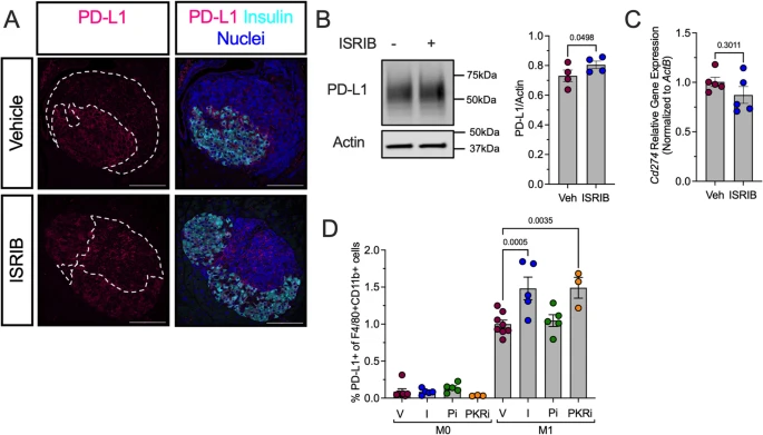
Fig. 8 ISR inhibition increases PD-L1 protein levels in prediabetic female NOD mice. Prediabetic 6-week-old female NOD mice were treated with ISRIB (2.5 mg/kg) for two weeks. A Representative images of pancreata from vehicle- or ISRIB-treated NOD mice stained for PD-L1 (magenta), insulin (cyan), and nuclei (blue). Dotted lines indicate insulitic regions. Scale bars: 100 μm. B Representative immunoblot showing PD-L1 and actin in M1-polarized BMDMs treated with vehicle or ISRIB (left panels) and quantification of PD-L1 levels (right panel); n = 4 biological replicates. C Relative Cd274 mRNA levels measured by qRT-PCR normalized to Actb in M1-polarized BMDMs in the presence or absence of ISRIB; n = 5 biological replicates. D Flow cytometry analysis of PD-L1 + cells as a percentage of F4/80 + CD11b + BMDMs under M0 and M1 polarization conditions; n = 4–5 biological replicates. Statistical tests: Paired t-test: B and C, or 1-way ANOVA with Tukey post-hoc test: D. Data are represented as mean ± SEM