
|
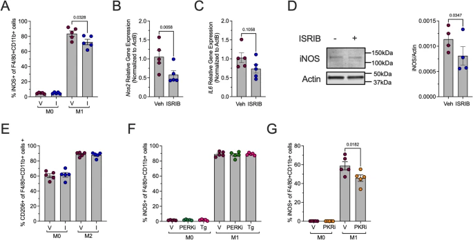
Fig. 4 ISR and PKR inhibition reduces proinflammatory macrophages. BMDMs from male 8-week-old C57Bl/6J mice were unpolarized (M0) or polarized to M1-like proinflammatory macrophages with and without 50 nM ISRIB. A Flow cytometry analysis of iNOS + cells as a percentage of F4/80 + CD11b + cells; n = 5 biological replicates. B Relative Nos2 mRNA levels measured by qRT-PCR normalized to Actb in M1-polarized BMDMs; n = 5 biological replicates. C Relative Il6 mRNA levels measured by qRT-PCR normalized to Actb in M1-polarized BMDMs; n = 5 biological replicates. D Representative immunoblot showing iNOS and actin in M1-polarized BMDMs (left panel) and quantification of iNOS levels; n = 4 biological replicates. E Flow cytometry analysis of CD206 + cells as a percentage of F4/80 + CD11b + cells; n = 5 biological replicates. F Flow cytometry analysis of iNOS+ (M1-polarization marker) cells as a percentage of F4/80 + CD11b + cells after pretreatment with PERK inhibitor (HC-5770; PERKi) or thapsigargin (Tg); n = 5 biological replicates. G Flow cytometry analysis of iNOS+ (M1-polarization marker) cells as a percentage of F4/80 + CD11b + cells after pretreatment with PKR inhibitor (C16; PKRi); n = 5 biological replicates