
|
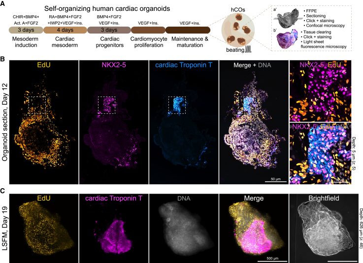
Figure 7
DNA replication in self-organizing 3D human cardiac organoids
(A) Protocol outline to generate self-organizing and beating hiPSC-derived human cardiac organoids (hCO), showing two distinct imaging strategies: FFPE organoid confocal imaging (a’) and 3D light sheet microscopy (b’).
(B) FFPE-processed day 12 organoid and confocal tile imaging of EdU fluorescence in DNA replicating cells (
(C) Whole-mount light sheet microscopy (LSFM) 3D visualization of a hCO at day 19 showing EdU positive cells and cTnT expressing cells. DNA-labelled nuclei are also shown (