
Image
|
Figure Caption
Figure 3
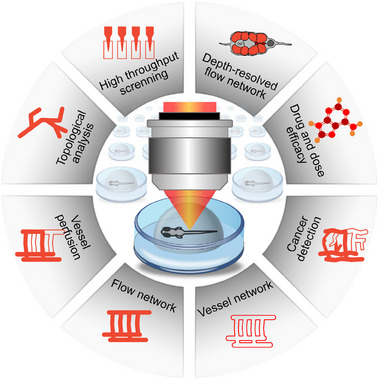
Overview of features that can be obtained in the context of vascular zebrafish imaging. The combination of OCT and fluorescence imaging allow high throughput screening for topological and vessel perfusion analysis, flow and vessel network visualization. Cancer development can be observed and compounds efficiency studies can be performed. By means of OCTA depth‐resolved flow network images are generated.
Acknowledgments
This image is the copyrighted work of the attributed author or publisher, and
ZFIN has permission only to display this image to its users.
Additional permissions should be obtained from the applicable author or publisher of the image.
Full text @ Adv Sci (Weinh)