
Fig. 5
- ID
- ZDB-IMAGE-250902-48
- Publication
- Sugitani et al., 2025 - The Rapid Activation of MYDGF Is Critical for Cell Survival in the Acute Phase of Retinal Regeneration in Fish
- All Figures
- Figures for Sugitani et al., 2025
|
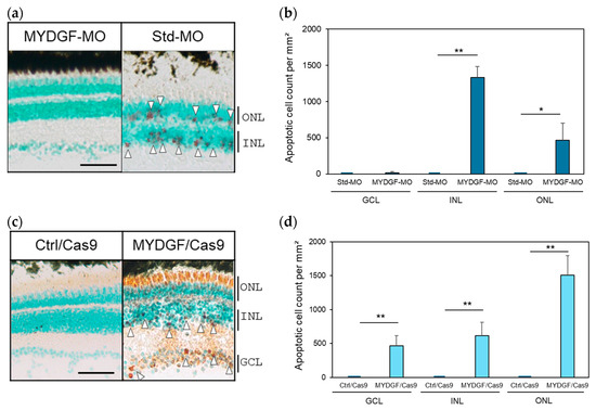
Fig. 5 Suppression of MYDGF expression after ONI increased the number of apoptotic cells in the retina and disrupted the retinal layer structure. (a) Injection of MYDGF-specific MO (MYDGF-MO) significantly increased the number of red-stained apoptotic cells 3 h after ONI (white arrowhead); in comparison, no apoptotic cells were observed in the control-MO (Std-MO) injection group. (b) Apoptotic cells were quantified as the number of apoptotic-positive cells per mm2 in the retina. (c) Administration of MYDGF-specific sgRNA via the CRISPR/Cas9 system (MYDGF/Cas9) also significantly increased the number of red-stained apoptotic cells (white arrowhead), and most of the photoreceptors were stained red in the ONLs. No apoptotic cells were observed in the control/Cas9 injection group (Ctrl/Cas9). (d) Apoptotic cells in the retina were quantified as the number of apoptotic cells per mm2 following CRISPR/Cas9 system administration. Each nuclear layer of the retina was stained with 3% methyl green for counterstaining. Data are presented as the mean ± SEM (n = 5–6) of three independent experiments and were analyzed using one-way ANOVA. Statistical significance was set at ** p < 0.01 and * p < 0.05. ONL: outer nuclear layer; INL: inner nuclear layer; GCL: ganglion cell layer. Scale bar = 50 µm