
|
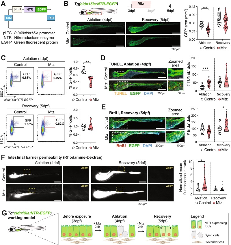
Fig. 1 Loss and recovery of GFP+ IECs after Mtz treatments of the Tg(cldn15la:NTR-EGFP) reporter. (A) Generated plasmid containing the 0.349cldn15la promoter upstream Nitroreductase (NTR) and EGFP fusion protein. (B) Changes in the GFP+ intestinal area during Mtz-induced ablation (4dpf) and recovery (5dpf). Scale bar = 200 µm. Pooled data from 3 independent experiments (1 dot = 1 larva). (C) Whole-larva flow cytometry analysis of control and Mtz-treated larvae during ablation and regeneration. Each dot represents 1 independent experiment (1 dot = pool of 10 larvae, 5 independent experiments conducted). (D) Apoptosis analyses by whole-mount terminal deoxynucleotidyl transferase dUTP nick end labeling (TUNEL) of Mtz-treated larvae. Scale bar = 200 µm in whole-intestine pictures and 50 µm in zoomed area. Pooled data from 2 independent experiments (1 dot = 1 larva). (E) 5-bromo-2′-deoxyurinide (BrdU) incorporation in Mtz-treated larvae. Scale bar = 200 µm in whole-intestine pictures and 100 µm in zoomed area. Pooled data from 2 independent experiments (1 dot = 1 larva). (F) Intestinal permeability of control and Mtz-treated larvae assessed by oral gavage of rhodamine-dextran. Normalized mean fluorescence in the trunk was measured. Scale bar = 200 µm. Pooled data from 2 independent experiments (1 dot = 1 larva). (G) Working model. Different symbol shapes denote independent experiments in B, C, D, E, and F. Unpaired t-tests were performed in B, C, D, E and F to compare conditions at specified timepoints. *p < 0.05; ***p < 0.001.