IMAGE

Fig. 8.

ID
ZDB-IMAGE-250828-149
Source
Figures for Yu et al., 2025
Image
Figure Caption

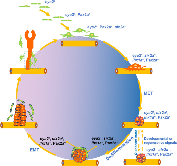
Fig. 8. Schematic overview of zebrafish RSC renewal and differentiation.

During zebrafish embryonic development, RSCs arise from eya2+ mesenchymal cells (shown as green cells). These cells then express Pax2a and six2a, maturing into functional RSCs. By 8 dpf, they begin expressing lhx1a (indicated as red cells) and aggregate, undergoing a MET to form compact cell aggregates. In the absence of developmental or injury-related signals, these aggregates enter a quiescent state. Upon reappearance of such signals, the aggregates reactivate: Base cells differentiate into nephrons, while apex cells regenerate RSCs through dedifferentiation and EMT. The newly formed RSCs reenter a cycle of generating new nephrons and additional RSCs.

Acknowledgments
This image is the copyrighted work of the attributed author or publisher, and ZFIN has permission only to display this image to its users. Additional permissions should be obtained from the applicable author or publisher of the image. Full text @ Sci Adv