IMAGE

Figure 1

ID
ZDB-IMAGE-250816-74
Source
Figures for Eckert et al., 2025
Image
Figure Caption

Figure 1

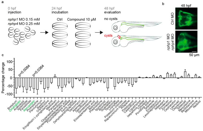
Drug repurposing screen to identify the compounds affecting cyst formation in a zebrafish nephronophthisis model. (a) Experimental workflow of the drug repurposing screen. Zebrafish oocytes were injected with MO targeting nphp1 (0.15 mM) and nphp4 (0.25 mM). At 24 hpf, groups of 20 embryos in triplicate were treated with either 10 µM of test compound or DMSO control. Phenotypic evaluation was performed at 48 hpf, assessing for glomerular cysts (red arrows), a characteristic feature of nephronophthisis. (b) Representative fluorescence microscopy images of zebrafish pronephric glomeruli at 48 hpf. The top panel shows normal glomerular morphology in Ctrl MO-injected embryos. Bottom panel demonstrates glomerular cyst formation in nphp1/nphp4 double-morphant embryos. Scale bar: 50 µm. (c) Quantification of the percentage change in cyst formation compared to DMSO controls for all tested compounds. Compounds that were further characterized are highlighted in green (Omarigliptin, Atorvastatin). Data are presented as mean ± SEM.

Acknowledgments
This image is the copyrighted work of the attributed author or publisher, and ZFIN has permission only to display this image to its users. Additional permissions should be obtained from the applicable author or publisher of the image. Full text @ Int. J. Mol. Sci.