IMAGE

Fig. 2

ID
ZDB-IMAGE-250816-12
Source
Figures for Yamashita et al., 2025
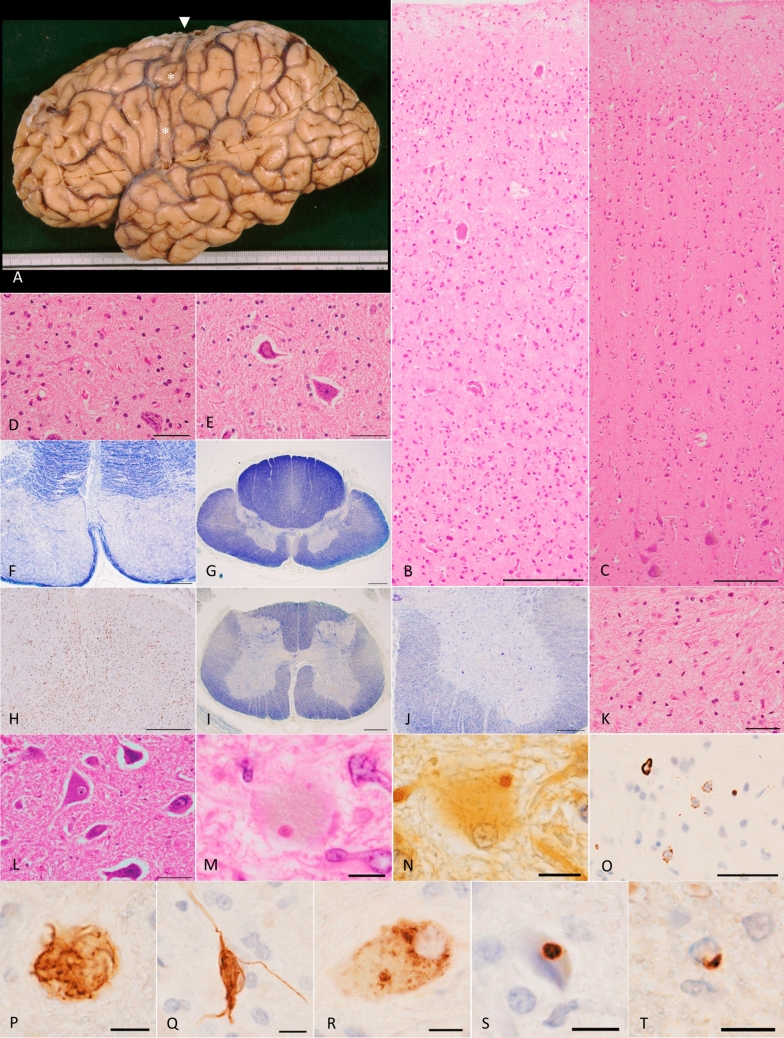
Image
Figure Caption

Fig. 2

Phosphorylated TDP-43 pathology, Bunina bodies, and severe loss of the upper and lower motor neurons in the older brother (IV-1) with a homozygous c.518dupC frameshift variant in DNAJC7.a Lateral view of the left hemisphere, showing severe atrophy in the precentral gyrus (asterisks). Arrowhead indicates the top of the precentral gyrus, the width of which is extremely thin. b Severe neuronal loss with gliosis in all cortical layers of the primary motor cortex. Betz cells are nearly completely absent. H&E stain. Scale bar: 200 μm. c Primary motor cortex in a 68-year-old pathologically normal control case. Betz cells are present in layer V, and the structure of the cerebral cortex is spared. H&E stain. Scale bar: 200 μm. d Severe neuronal loss with gliosis in the hypoglossal nucleus. A remaining neuron shows shrinkage of the cell body. H&E stain. Scale bar: 50 μm. e Hypoglossal nucleus in a 51-year-old pathologically normal control case. Neither glial proliferation nor shrinkage of neurons is observed. H&E stain. Scale bar: 50 μm. f Severe degeneration in the pyramidal tract within the medulla oblongata, as visualized by Klüver–Barrera stain. Scale bar: 500 μm. g, h Severe loss of myelin (g) with microglial activation (h) in the lateral tract of the thoracic cord. The dorsal spinocerebellar tract does not exhibit these changes. g Klüver–Barrera stain, h Iba-1 immunohistochemistry. Scale bars: g 1 mm and h 500 μm. i Degeneration in the lateral tract of the lumbar cord, as shown by Klüver–Barrera stain. Scale bar: 1 mm. j Severe loss of anterior horn cells in the lumbar cord, visualized by Klüver–Barrera stain. Scale bar: 500 μm. k Severe gliosis with loss of neurons in the anterior horn of the lumbar cord. H&E stain. Scale bar: 50 μm. l The Spinal anterior horns in a 67-year-old pathologically normal control case. Neither loss of anterior horn cells nor gliosis is present. H&E stain. Scale bar: 50 μm. m Bunina bodies in the lumber anterior horn. H&E stain. Scale bar: 10 μm. n Bunina body in the lumbar anterior horn, identified by Cystatin C immunohistochemistry. Scale bar: 10 μm. o Phosphorylated TDP-43-positive NCIs in the upper layers of the primary motor cortex. Neurites are barely discernible. pS409/410-2 immunohistochemistry. Scale bar: 50 μm. ps Various morphologies of TDP-43-positive NCIs in the lumbar anterior horns. Inclusions include p skein-like, q neurofibrillary change-like, r diffuse granular, and s round structures. pS409/410-2 immunohistochemistry. Scale bars: 10 μm. t Phosphorylated TDP-43-positive glial cytoplasmic inclusion in the pyramidal tract of the medulla oblongata. pS409/410-2 immunohistochemistry. Scale bar: 10 μm. c, e, l Control cases presented here lacked any neurodegenerative changes except for minimal NFTs with Braak stage I. H&E, hematoxylin–eosin; NCI, neuronal cytoplasmic inclusions; NFTs, neurofibrillary tangles; TDP-43, TAR DNA-binding protein of 43 kDa

Acknowledgments
This image is the copyrighted work of the attributed author or publisher, and ZFIN has permission only to display this image to its users. Additional permissions should be obtained from the applicable author or publisher of the image. Full text @ Acta Neuropathol.