
|
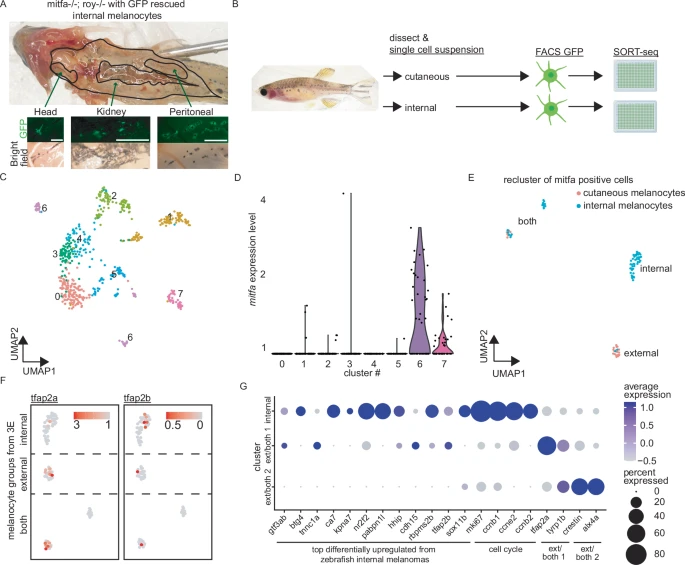
Fig. 3 Zebrafish internal melanocytes have properties consistent with MM initiating cells. A Light micrographs demonstrating internal GFP-expressing melanocytes from adult Casper zebrafish. Scale bar = 500 μm. n = 7 zebrafish. B Schematic for single-cell RNA-seq (scRNA-seq) of cutaneous and internal adult zebrafish melanocytes. Cutaneous melanocytes were combined from two zebrafish and mucosal melanocytes were combined from seven zebrafish. Created in BioRender. Insco, M. (2025) https://BioRender.com/da5ul5n. C UMAP of scRNA-seq from GFP-sorted external and internal melanocytes. D mitfa-expressing cells from (C). E UMAP of sub-clustered mitfa+ melanocytes. Blue = from internal. Red = from cutaneous. F tfap2a and tfap2b expression in “internal”, “external”, and “both” melanocyte groups from (E). G Dot plot showing gene expression of top differentially expressed genes from zebrafish MMs for Seurat-defined melanocyte clusters from Fig. S3D.