
|
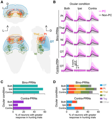
Fig. 5 Bino-PRNs are motor modulated and receive binocular input in no behavior trials (A) Anatomical locations of bino-PRNs in four selected regions of the brain; coronal, horizontal, and sagittal views. (B) Average responses of bino-PRNs in each region and mono-contra neurons in the OT to PC and non-PC trials (defined by eye convergence) to prey at 0° in different ocular conditions. In the ipsi condition, only the ipsilateral eye relative to the PRN was able to see the stimulus. Solid lines and shaded regions represent mean and ±1 SD, respectively. (C) Percent of bino- and contra-PRNs with significantly larger responses during hunting trials. Trial response is calculated as the mean dF/F between +1 and +3 s after stimulus onset. A neuron’s response is considered significantly greater if the one-sided Mann-Whitney U test, comparing hunting trial responses to NR trial responses, results in a p value less than 0.05. (D) Proportion of bino- and contra-PRNs with significantly greater responses in hunting trials in each brain area. See also Figure S5.